We would like to invite members of the drug discovery community to learn about special considerations for 3D Spheroid and Organoid Imaging. Together with experts at InSphero and Yokogawa, we are hosting a webinar on January 14, 2021. In this webinar, we share our experience and advice for sample preparation, imaging, and analysis for both fixed and live-cell in high content screening assays. We hope to provide the research community with tips and tricks for 3D drug screening, highlighting practical aspects of drug screening in discovery and therapy with 3d multicellular models.
High content imaging for drug screening
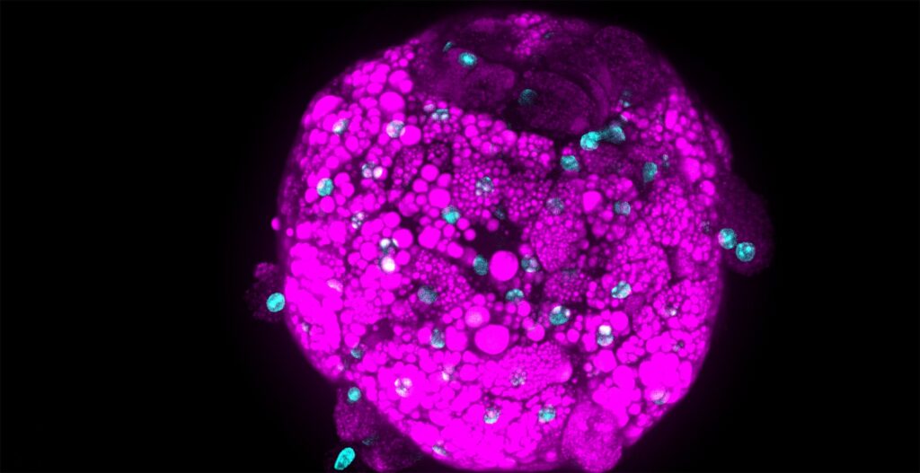
Conventional drug screening has mostly been limited to 2d cell cultures in multi-well plates. The 2D cell culture model is being replaced with 3D models as a more physiologically relevant in vitro model choice. The behavior of cells differs in three-dimensional cell contacts. Tissue relevant co-cultures and integrated signaling, along with o2/co2, nutrient, waste, and drug gradients are quite different in 2D vs 3d. High content imaging enables clinically relevant interrogation of 3d models, along with targeting of specific cell-cell interactions of target and non-target cell types. Using spinning disk confocal microscopy allows long-term imaging of live samples due to low phototoxicity and optical sectioning over 100um deep into 3d tissue samples.
With careful choice of instruments and sample holders, even month-long longitudinal studies of live samples are possible. We will show some examples of how live imaging vs fixed imaging can change regarding throughput, kinetic data, probes, and signal strength.
Looking at cell level data
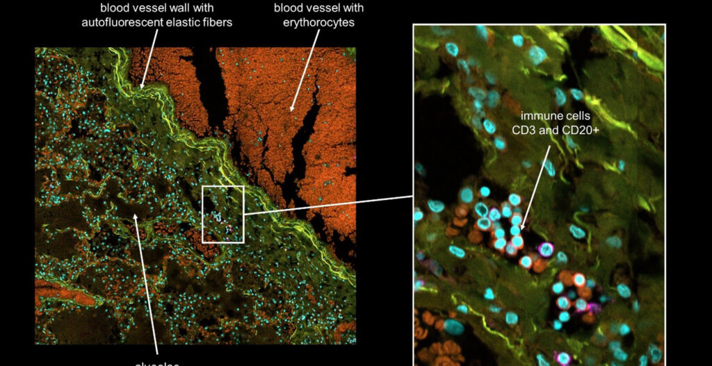
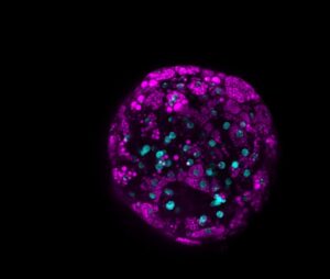 It is important to keep in mind: probe and antibody penetration kinetics, taking into consideration sample fixation and permeabilization conditions, along with incubation time, and any non-specific binding that may occur. For fixed tissue, tissue clearing can be an excellent choice to increase light penetration allowing for a larger sample size, which can be important to understand the larger picture of cell-cell interactions. By looking at cell level data, we can count cells, look for intracellular biomarker expression, individual cell fate populations, along any intercellular structures. The spatial information along with cell infiltration and migration or signaling in defined zones due to kinetic changes in cell number or biomarker expression can be achieved in live sample preparation.
How to understand the complex volume of data
We will discuss common ways to report results of analysis, including heatmaps, dose-response curves, and per well pie charts. We will include a brief discussion of Machine Learning and Deep learning tools for segmentation and phenotypic assignment. These automated tools can help researchers to understand the complex volume of data in these high content imaging analysis systems.
I’m looking forward to an exciting discussion. Will I see you there?
[vc_cta h2=”On-demand Webinar” h4=”Optimizing High-Content of 3D Models for Drug Discovery” color=”white” add_button=”bottom” btn_title=”Register” btn_color=”warning” btn_link=”url:https%3A%2F%2Fwww.labroots.com%2Fms%2Fwebinar%2Foptimizing-high-content-imaging-3d-models-drug-discovery-2%3Futm_source%3Dblog%26utm_medium%3Dbanner%26utm_campaign%3Dyca_webinar_en|title:on-demand%20webinar|target:_blank” css=”.vc_custom_1616607925302{background-color: #004f9b !important;}”]Click here for watching the webinar on-demand![/vc_cta]