Advanced HCA (High Content Analysis) platform with multicellular 3D models for drug discovery
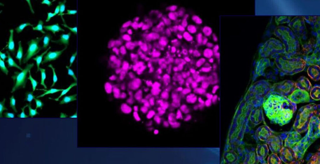
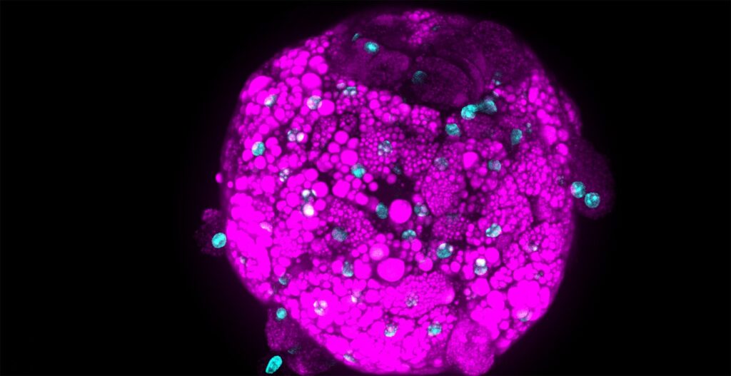
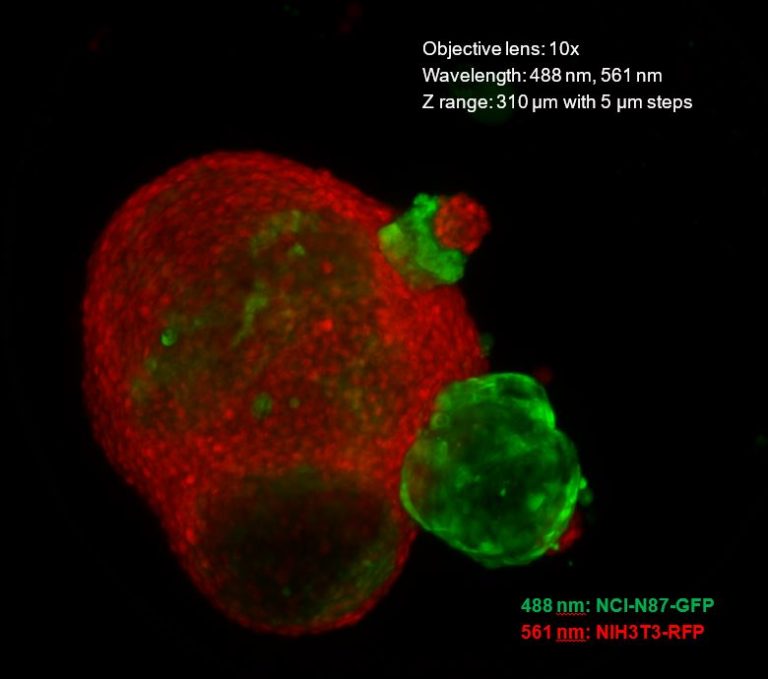
InSphero AG, a biotech based in Schlieren, Switzerland, develops 3D cell-based platforms for drug development and safety testing and sets the gold standard for advanced, yet easy-to-use 3D assay solutions for predictive compound classification. The company’s 3D InSight™ microtissues are complex multicellular models with a 3D architecture that enables the formation of cell-to-cell contacts and distinct microenvironment characteristics of native human tissues, making them ideal for preclinical testing of compounds of interest.
InSphero and their pharma and biotech partners were eager to exploit the full potential of 3D models through interrogation of specific cell population interactions and extraction of spatial and temporal information. High Content Analysis (HCA), specifically high content imaging, was recognized as a key technology to accomplish these technically challenging goals.
High-resolution 3D imaging
In support of that goal, InSphero recently introduced the Akura™ 384 plate, a 384-well microplate optimized for 3D cell culture and HCA. While planning the launch of this new plate, InSphero began actively seeking HCA partners offering platforms with high-resolution 3D imaging and 3D imaging analysis capabilities to help evaluate the full potential of the combined technologies (3D cell models and HCA). Frauke Greve, InSphero Product Manager for Cell Culture Consumables, researched the HCA market and contacted the Yokogawa Life Innovation Business team based in Kanazawa, Japan.
For watching the video from InSphero, simply click on the picture below.

High Content Analysis: Microscopic images at high speed
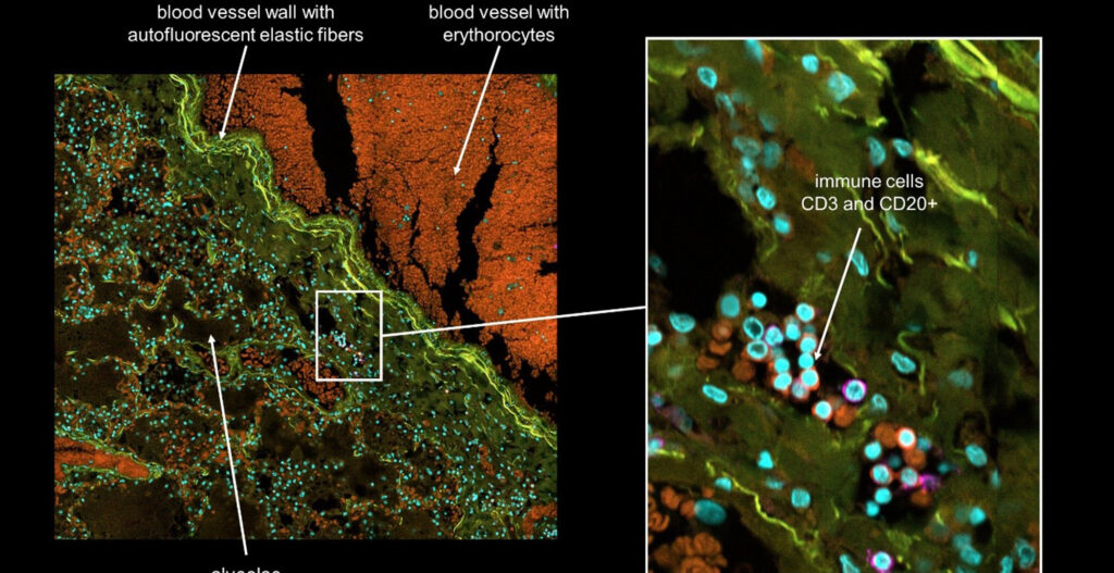
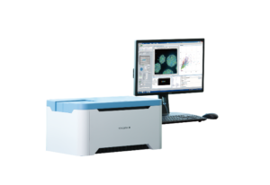
Yokogawa’s range of HCA product portfolio include CellVoyager CV8000, which is a high-end system, CQ1 (Picture 1), which is a benchtop instrument, and CellPathfinder™ analysis software suite. By using confocal scanning technologies based on microlens-enhanced Nipkow disks, Yokogawa HCA instruments automatically acquire a large amount of high-resolution, 2D/3D confocal microscopic images at high speed with minimal photo-toxicity and photo-bleaching. Furthermore, the information contained in acquired images can be quantified by linking individual data points in graphs to their corresponding microscopic image. Yokogawa HCA systems also provide ideal culture conditions for live cells through temperature control, CO2 control, and humidification. These functions allow monitoring of dynamic physiological changes at the cellular level.
“We are always looking for collaborations with life scientists, who would like to utilize our imaging technologies and solutions in their research. We were glad InSphero reached out to us,” said Mahomi Suzuki, Application Scientist from Yokogawa. The first step in this new collaboration between InSphero and Yokogawa was to perform imaging trials. In January 2019, the first tests were conducted at the Yokogawa research laboratories in Kanazawa, Japan. Samples were run on two Yokogawa CellVoyager instruments: The CQ1 benchtop Confocal Quantitative Image Cytometer and the CV8000 High-throughput Cytological Discovery System. The CQ1 instrument acquisition conditions were very compatible with InSphero’s Akura™ 384 plates. Therefore, the CQ1 instrument was selected as a perfect fit for the targeted research applications.
Several weeks planning studies and preparing samples
After the initial test phase in Japan, InSphero spent several weeks planning studies and preparing samples for an in-house research project with the CQ1 instrument. In August 2019, with the support of Cenibra GmbH a CQ1 system was placed at InSphero AG in Schlieren, Switzerland, for the collaboration project. After the CQ1 was installed at InSphero, there was one month of intense collaboration during which InSphero scientists and Yokogawa technical support scientists worked side-by-side to acquire high-quality images from a wide array of experiments. The InSphero team screened many different tissue and disease models with dozens of fluorescent endpoints during the month-long research project.
Their studies included fixed and live-cell studies, 3D and 4D analysis (3D plus time-series). The high level of enthusiasm for access to the CQ1 imager often created bottlenecks at the instrument. It was especially challenging for InSphero’s team to schedule all their 3D fixed cell studies with the 4D live-cell imaging studies as the latter often occupied the instrument for several days at a time. However, the team found that onsite access to the CQ1 imager helped foster a heightened appreciation of the potential for HCA. There was a general consensus among the team that this research collaboration only “touched the tip of the iceberg” with regards to the true potential of combining multicellular 3D models with high-resolution 3D imaging. By the end of August, many new ideas were generated, some of which could not be tested due to time constraints.
High content analysis: Image acquisition
During the research collaboration, it became clear that generating samples appropriate for imaging can involve numerous rounds of optimization. Both image acquisition and analysis can be challenging. Neither can be mastered in a few weeks. Therefore, ongoing support from the instrument provider is an essential element for success. A comprehensive toolset of 3D image and data analysis tools are necessary for 3D models as different biological applications require different 3D image analysis approaches.
In October 2019, InSphero was invited by guest editors at the journal SLAS Discovery to submit a paper for an upcoming special issue on HCA. This invitation came at a particularly opportune time just after completion of the research project with Yokogawa. A decision to use the Yokogawa imaging data for this manuscript resulted in a further extension of the InSphero-Yokogawa collaboration. At the end of 2019, the project teams at both companies contributed to image analysis and data visualizations for the final manuscript. The HCA data and results presented in ‘Results and Discussion’ section were generated using InSphero 3D multi-cellular models with Yokogawa CQ1 instrument and CellPathfinder™ software suite. As of this writing, the paper has been conditionally accepted and is expected to be published in July or August 2020.
The citation for this paper is:
- Wardwell-Swanson, J., Suzuki, M., et al. (In Press) A Framework for Optimizing High Content Imaging of 3D Models for Drug Discovery. (Manuscript conditionally accepted by SLAS Discovery for publication.)
Part of the research results included in this paper were also presented during the Society of Laboratory Automation and Screening annual meeting held in January 2020 in San Diego:
- Wardwell-Swanson, J., Suzuki, M., et al. (2020) A Framework for Optimizing High Content Imaging of 3D Models for Drug Discovery. (SLAS poster)
- Lichtenberg, J., Frey, O., Suzuki, M., Why the Akura™ Technology Platform Enables a Seamless Link from Screening in 3D to Organ-on-a-Chip. (Exhibitor tutorial presentation)
Combination of MapMode targeted acquisition and large-format sCMOS camera
From a technical standpoint, researchers at InSphero learned that conducting 72 hours of live-cell 3D imaging without noticeable phototoxicity is absolutely possible. This represents a remarkable breakthrough in HCA systems and proves the quality engineering of Yokogawa’s enclosed environmental chamber, sCMOS camera, dual spinning disk technology, and adjustable laser power. The InSphero team also learned that the combination of MapMode targeted acquisition and large-format sCMOS camera enables the acquisition of a 250 µm spheroid in a single field with a 20x objective. They determined that was a significant technical and time-saving improvement over a CCD camera with non-targeted acquisition, which often results in the acquisition of a large number of empty fields and requires image stitching to join adjacent fields containing parts of the object of interest.
A great partnership
InSphero and Yokogawa scientists concurred that there are interesting and important biological questions (e.g., cell-to-cell interactions, cell-population responses, and spatial and temporal information) that can be best addressed in physiologically relevant 3D models using HCA. There is already evidence that this research work is not just an InSphero-Yokogawa story, but the results of this technology partnership have engendered further interest and collaborations with other top pharma partners.
“One of our challenges as a biotech pioneer is to identify the right technology partners that help us deliver truly innovative solutions for drug discovery and safety testing. Our scientists and bioengineers set the bar high for Yokogawa. And they consistently exceeded our expectations. Both in terms of imaging and data analysis quality as well as the dedicated, enthusiastic support of their Life Science team. Together, I have no doubt that we will set new standards for high content analysis of 3D spheroids.”
This summarizes Jan Lichtenberg, Ph.D., InSphero CEO, and Co-founder.