Why Chromosome Folding Matters in Cell Biology
Understanding how chromosomes fold during mitosis is one of the most fundamental challenges in cell biology. Chromosome architecture determines how genetic material is organized, replicated, and segregated during cell division—a process essential for life and with implications in cell death, rare developmental disorders or cancer formation.
Recently, scientists at the European Molecular Biology Laboratory (EMBL) published a new study that illuminates this complex process on high-resolution level for the first time. Their research, detailed in Cell, introduces new insights into the mechanisms of chromosome folding during mitosis.
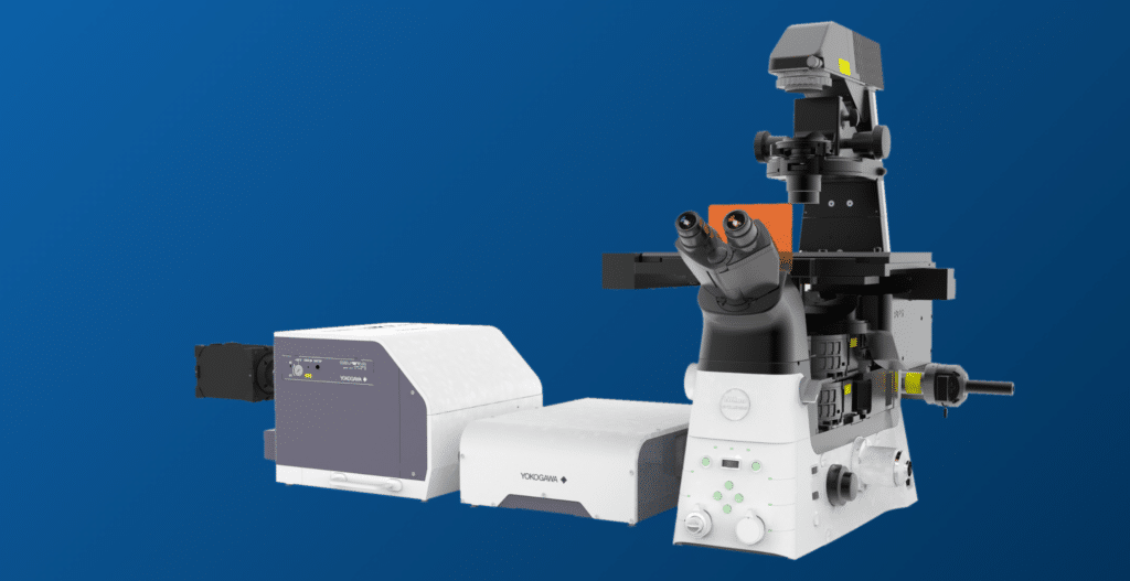
Achieving these results required more than scientific ingenuity—it demanded imaging technology capable of capturing nanoscale chromatin structures in living cells with unprecedented clarity. This is where Yokogawa’s CSU-W1 SoRa confocal system, combined with Nikon’s advanced ECLIPSE Ti2-E microscope platform, played a pivotal role.
EMBL’s Breakthrough: 3D Chromatin Tracing
The EMBL team tackled a long-standing challenge: visualizing the dynamic folding of chromosomes in three dimensions during cell division. Traditional imaging methods often fall short, either lacking the resolution to resolve fine chromatin structures or introducing phototoxicity that compromises live-cell observations.
By applying a novel nanoscale 3D chromatin tracing approach called Loop trace, researchers mapped chromosome architecture at resolutions previously unattainable in living cells. This technique revealed how chromosomes compact and organize themselves during mitosis—forming very large and overlapping loops mediated by protein complexes called Condensins. Building these loops, the chromosomes get compacted and kept in form.
These findings not only deepen our understanding of genome organization but also open new avenues for studying how disruptions in these processes contribute to disease. For a detailed look at the methodology, Nikon has published an application note explaining the technical underpinnings of this breakthrough.
The Technology Behind the Discovery
How CSU-W1 SoRa Enables Nanoscale Live-Cell Imaging
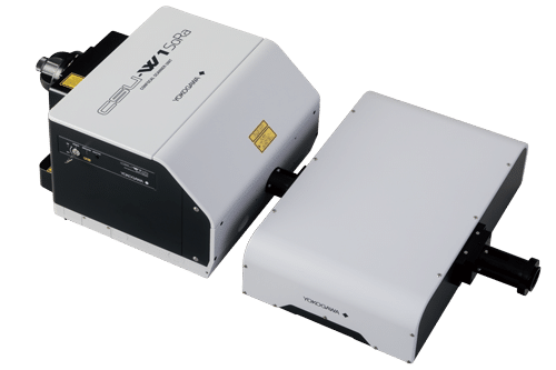
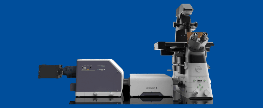
At the heart of this achievement lies Yokogawa’s CSU-W1 SoRa—a super-resolution spinning disk confocal system designed for live-cell imaging. SoRa combines the speed and low phototoxicity of spinning disk confocal microscopy with optical super-resolution, enabling researchers to visualize structures down to ~120 nm without compromising cell viability.
This capability was essential for capturing the fine details of chromatin organization during mitosis. The CSU-W1 SoRa allowed the EMBL team to perform high-speed, high-resolution imaging over extended periods, preserving the integrity of living cells while revealing nanoscale features.
Nikon’s ECLIPSE Ti2‑E: An inverted microscope for a stable, high-performance platform
Complementing this system was Nikon’s ECLIPSE Ti2-E inverted microscope with Perfect Focus System 4 (PFS 4), which provided a stable, high-performance platform for integration with Yokogawa’s confocal technology. In combination with Nikon’s high‑quality objectives, the setup offered the optical precision needed for demanding live‑cell and high‑content imaging workflows. When paired with fully automated, customized microfluidics, enabled through the JOBS module in Nikon’s NIS‑Elements software, the system delivered a robust, end‑to‑end imaging solution. Together, these instruments formed a powerful imaging solution that made nanoscale 3D chromatin tracing possible.
A Testament to Collaboration
This research exemplifies what can be achieved through strategic collaboration. Yokogawa and Nikon have a long-standing partnership focused on advancing imaging technologies for life sciences. By combining Yokogawa’s expertise in confocal systems with Nikon’s leadership in optical microscopy, the two companies have enabled scientists to push the boundaries of what is observable.
The EMBL study stands as a flagship example of this synergy. It demonstrates how joint innovation can empower researchers to tackle complex biological questions and deliver insights that were once out of reach.
Implications for Cancer Research, Developmental Biology, and Drug Discovery
The ability to visualize chromosome folding at nanoscale resolution in living cells has far-reaching implications:
– Cancer Research: Chromosome misfolding and segregation errors are hallmarks of cancer. Understanding these processes at a structural level could inform new diagnostic and therapeutic strategies.
– Developmental Biology: Chromatin architecture influences gene expression during development. High-resolution imaging can help unravel how these patterns are established and maintained.
– Drug Discovery: By revealing how chromosomes respond to chemical perturbations, advanced imaging can accelerate the development of targeted therapies.
As imaging technologies continue to evolve, Yokogawa remains committed to supporting life science innovation. Our goal is to provide researchers with tools that not only meet today’s challenges but also anticipate tomorrow’s discoveries. The EMBL study is more than a scientific milestone—it is a testament to the power of technology and collaboration. By combining Yokogawa’s CSU-W1 SoRa with Nikon’s advanced microscopy platform, researchers have unlocked new insights into the architecture of life at its most fundamental level.
To learn more, explore these resources:
– EMBL Research Story: How chromosomes shape up for cell division | EMBL
– Cell Journal Paper: Nanoscale DNA tracing reveals the self-organization mechanism of mitotic chromosomes: Cell
– Nikon Application Note: Nanoscale 3D chromatin tracing using the Nikon ECLIPSE Ti2-E with Yokogawa CSU-W1 SoRa spinning disk system | Application Notes | Hintergrundwissen | Nikon Europe B.V.
– Yokogawa CSU-W1 SoRa: CSU-W1 SoRa Confocal Scanner Unit | Yokogawa Europe