The Yokogawa Life Innovation Business expands towards Single-Cell Analysis (SCA)
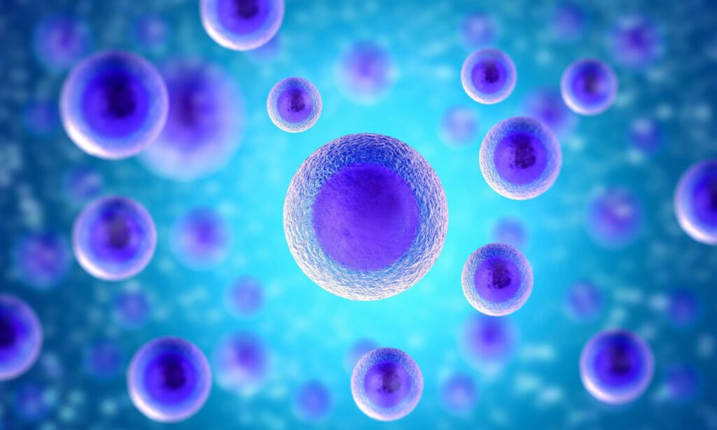
When people think about a cell population most envision a more or less evenly distributed layer of equal cells. However, tissues of organisms are composed of different cell types of which some occur only in small numbers. For example, stem cells or metastasis in tumor diseases. Also, when performing experiments in the lab there are always some cells that are behaving differently than their “sibling cells”. Additionally, we all know that severe diseases like cancer can originate from a single cell that is somewhat different respectively contains errors in its genes.
Gaining insights
Having that said, gaining insights into a single cell versus a whole cell population is important for studying mechanisms of programmed cell death (Apoptosis) or proliferation as well as the migration of cells. With Single-Cell Analysis (SCA) researchers hope to better understand the onset of diseases and mechanisms of cell signaling.
Patch-clamp techniques are used for example to study the electrophysiology of cells like neurons. It is also interesting to collect DNA, RNA, or protein samples from single cells for further analysis. This is possible via mass spectroscopy, RT-PCR, or DNA sequencing. In 2013, the Nature Methods journal selected DNA and RNA sequencing of single cells as the method of the year1.
New Solutions for Single-Cell Analysis
The Yokogawa Life Innovation Business recently developed in-house and through the acquisition of BioStinger Inc. new solutions for Single-Cell Analysis (SCA). Today we would like to briefly introduce two new SCA research solutions. These products are expecting to be commercially available in the European market in the next months.
Single Cellome™ Unit SU10
[vc_single_image image=”663″ img_size=”large” alignment=”right”]
The first new product to modify single living cells is the minimally-invasive intracellular nano-injector named Single Cellome™ Unit SU10. The Single Cellome Unit can be mounted to an existing inverted microscope. It uses a nano-scale pipette tip [nm] to penetrate cells to either inject a substance into or aspirate content from a single target cell. The procedure is of low invasiveness. It allows live-cell imaging of single cells after substance injection into the nucleus or cytoplasm. The low invasiveness is achieved by using a glass pipette with a tip diameter of approximately 100 nm (nano-scale). The tip movement speed is automatically controlled to minimize damage to the cell during penetration and retraction of the nanopipette.
Accurate positioning at and penetration of the target single cell is performed by a high-performance 3-axis actuator (XYZ directions). The automated Z-direction movement detecting the cell surface as well as controlling the penetration and retraction of the nanopipette tip is based on continuous ion current measurement.
For which application cases can you use the SU10?
Direct injection of substances such as vector and genome editing tools (CRISPR/Cas9) into the nucleus of a target single cell could be considered. Furthermore, efficacy and toxicity evaluation of potential drug candidates could be studied in a single cell of interest. In the Yokogawa development laboratories in Japan, single-cell injection success rates of up to 95% were achieved by our researchers. Therefore, we believe the SU10 could be a valuable instrument to support genome engineering researchers in their daily work.
SS2000 Single-Cell Autosampler
A second product entering the market a few months after the SU10 will be the so-called SS2000 Single-Cell Autosampler. This SCA instrument is based on the Yokogawa bench-top CellVoyager CQ1. It uses the technical properties of the CQ1 system to image sample cells. Applying analysis features, cells of particular interest can be identified and selected by the researcher. Using a pipette with a tip in the micro-meter [µm] range those cells of interest are collected one by one in an automated fashion. Each cell is transported to a buffered solution where it is immediately frozen and stored for further analysis like PCR or mass spectroscopy. Additionally, a substantial amount of the cytoplasm containing organelles of interest for example identified by fluorescence labeling can be sampled, aliquoted, and stored in an automated fashion as well.
We are convinced both new instruments will be valuable and supportive to answer current research questions in the field of Single-Cell Analysis (SCA), gene editing, and drug discovery. Do you want to learn more about these fascinating technologies? Stay tuned!
[vc_cta h2=”Talk: Nanopipette Technology: A new tool for Single-Cell Analysis” color=”peacoc” add_button=”bottom” btn_title=”Watch now” btn_color=”warning” btn_link=”url:https%3A%2F%2Fbit.ly%2F3gzZF5i|title:Watch%20free%20on-demand%20webinar|target:_blank” css=”.vc_custom_1629986434084{background-color: #ffee00 !important;}”]Do you want to learn more about this promising technology?
Watch the On-Demand Webinar: Nanopipette Technology: A new tool for Single-Cell Analysis.
Speaker: Prof. Dr. Nader Pourmand (University of California Santa Cruz)
 [/vc_cta]