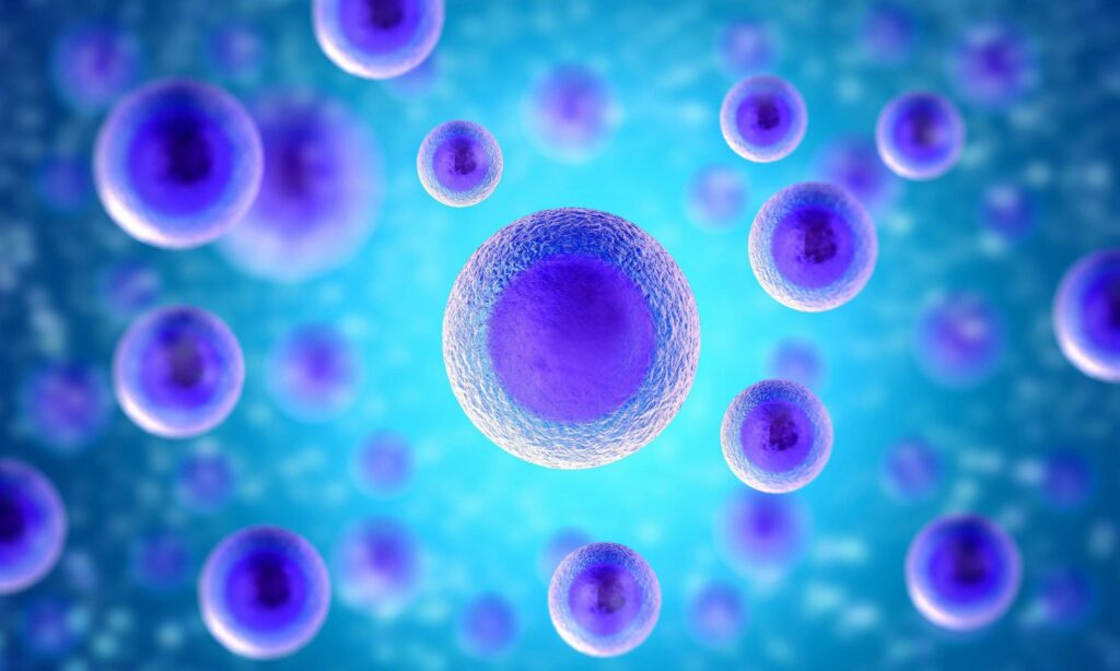
Spatial Transcriptomics is part of the -omics sciences and is defined as high-throughput quantification of gene expression, preferably highly multiplexed. This means that numerous genes are quantified at the same time within the same piece of tissue.
Omics technologies that are most frequently used include:
- Laser capture microdissection (LCM) which can be followed by RNA-sequencing for transcriptome-wide analysis
- Niche-seq.
- APEX-seq.
- single molecular fluorescent in situ hybridization (smFISH) based technologies like seqFISH
- MERFISH
- array-based technologies such as Spatial Transcriptomics (ST) or Visium
Transcriptomics: MERFISH
The most often used technology for single-cell transcriptome omics is MERFISH (Multiplexed Error-Robust Fluorescence in situ Hybridization) by vizgen.
MERFISH is a massively multiplexed single-molecule imaging technology. It measures simultaneously the copy number and spatial distribution of hundreds to tens of thousands of RNA species in individual cells. The disadvantage of hybridization technologies in omics sciences is the lack of detection of novel transcripts as compared to single-cell RNA sequencing. Whereas single-cell RNA sequencing loses spatial context during sample processing. Therefore, LCM followed by RNA-sequencing is best if novel transcript analysis in a spatial context is desirable.
Due to the laser that is used for LCM though, this method is very invasive compared to the automated sampling by our newly developed Single Cellome™ System SS2000.
Transcriptomics: GeoMX DSP
The platform GeoMX DSP from NanoString was developed for spatial omics of RNA or proteins. This method is based on the detection of protein or RNA by UV-photocleavable oligos that are bound to an antibody or RNAscope probes. The region of interest (ROI) can be imaged and chosen. After UV-cleavage of the oligos at the ROI, they are collected and sequenced apart from the omics.
Transcriptomics: Visium Platform
Another newly developed technology is the Visium platform from 10x Genomics. For this genomics technology, the sample has to be placed onto a Capture Area of the gene expression slide. Each Capture Area has thousands of barcoded spots containing millions of capture oligonucleotides with spatial barcodes unique to that spot. The samples can then be viewed with a fluorescence microscope.
For spatial omics with fresh frozen tissues, the tissue is permeabilized to release mRNA from the cells, which binds to the spatially barcoded oligonucleotides present on the spots. A reverse transcription reaction produces cDNA from the captured mRNA. The barcoded cDNA is then pooled for downstream processing to generate a sequencing-ready library. For omics with FFPE tissues, the tissue is permeabilized to release ligated probe pairs from the cells, which bind to the spatially barcoded oligonucleotides present on the spots. Spatial barcodes are added via an extension reaction. The barcoded molecules are then pooled for downstream processing to generate a sequencing-ready library.
The disadvantages of this spatial omics system are the lack of a single-cell resolution and the low detection efficiency.
How to succeed in single-cell spatial genomics
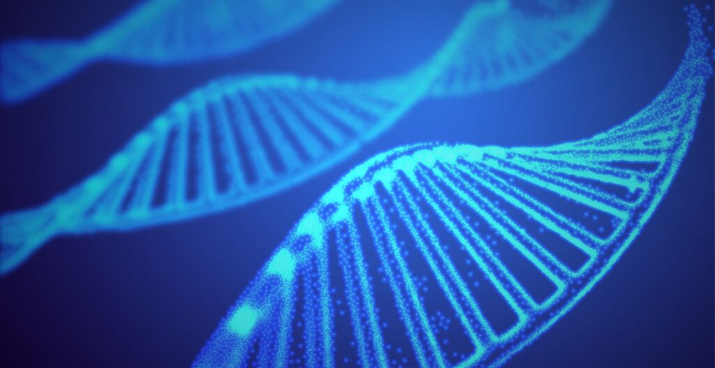
Single-cell spatial genomics relies on the ability the isolation of a single cell (for example by single-cell fluorescence-activated cell sorting (FACS), microfluidics), the amplification of its genome efficiently and accurately, and the sequencing of the DNA.
Several capture methods used for single-cell mono-omics analysis are commonly employed in single-cell multi-omics analysis, including low-throughput methods to capture tens or hundreds of cells, including laser capture microdissection and robotic micromanipulation, and high-throughput methods to capture tens of thousands of cells, including fluorescence-activated cell sorting (FACS) followed by plate-based isolation and the use of microfluidic platforms with microfluidic channels and reaction chambers or nanowells. Low-throughput spatial omics methods such as micromanipulation retain spatial information on the isolated cells, while this information is lost under high-throughput methods.
In contrast to the omics methods described above the automated single-cell sampler SS2000 can sample cells or intracellular components by retaining the spatial information for example in contrast to FACS in an automated manner without the need for special skills as it is for manual sampling.