
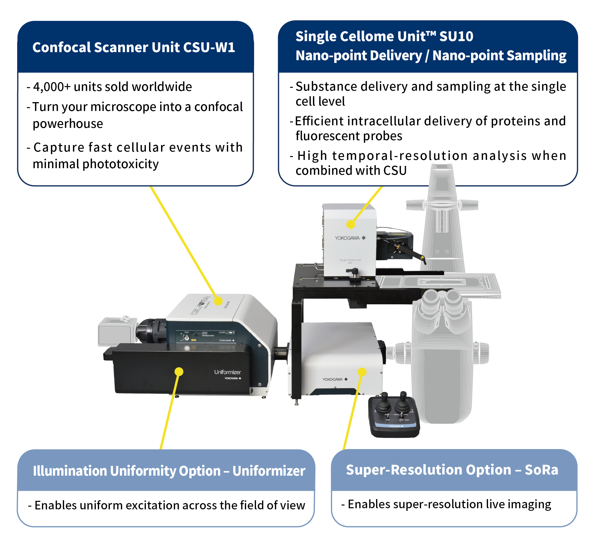
Single Cellome Unit SU10 enables the delivery of substances, such as recombinant proteins and genome editing tools, directly to the cytoplasm or nucleus of targeted single cells. It can also perform sampling at the single-cell level.
For use with an inverted optical microscope and a stereo microscope type. The microscope is not included with the SU10.
Single-cell Targeted Delivery & Sampling
- Supports research on liquid-liquid phase separation and organelles, such as mitochondria, autophagy, and proteasome
- Enables manipulation of phase separation by promoting or inhibiting droplet formation
- Enables directly targeting of the cytoplasm and nucleus
Efficient Intracellular Protein & Probe Delivery
- Fully compatible with live-cell imaging systems, such as the confocal scanner unit CSU series, enabling high temporal resolution analysis
- Delivers antibodies and fluorescent probes directly to the cytoplasm and nucleus
- Delivers genome editing tools such as ribonucleoproteins (RNPs)
Substance Delivery to Primary Cells and Plant Cells
- Enhances transfection efficiency in neurons
- Provides a promising new method for delivering substances into cells with rigid cell walls and high turgor pressure, such as plant cells
High-Efficiency, Low-Damage Intracellular Delivery
- Utilizes ultra-fine glass capillaries (nanopipettes) with tip diameters as small as several tens of nanometers
- Automates cell surface detection, membrane penetration, and material injection
Details
Application Examples
(1) Next-Gen Live-Cell Analysis : Precise Delivery with SU10, Dynamic Imaging with CSU*
| Intracellular delivery and structure formation of a phase-separating protein |
Monobody delivery for phase separation disruption |
Antibody delivery for intracellular molecular/organelle staining in live cells |
 |
 |
 |
| Cell : U2OS Substance : GFP-FUS |
Cell : HCT-116 Substance : Monobodies & Alexa Fluor™ 405-conjugated streptavidin |
Cell : HeLa Substance : Alexa Fluor™ 647- conjugated anti-TOMM20 antibody |
| Actin filament dynamics via peptide delivery in primary cells |
Intracellular environment measurement via nanoparticle delivery |
Intracellular dynamics of delivered substances |
 |
 |
 |
| Cell : Fish epithelial keratocyte Substance : Alexa Fluor™ 488- conjugated Phalloidin |
Cell : HeLa Substance : Qtracker 655 |
Cell : HeLa Substance : FITC-conjugated dextran |
*Yokogawa’s CSU confocal unit, featuring a proprietary dual Nipkow disk, is ideal for live cell imaging. Its micro lenses enhance excitation efficiency for high-speed imaging with minimal cellular toxicity and photobleaching. With over 4,000 units sold globally, it’s the industry standard for live cell imaging. Click here for CSU website.
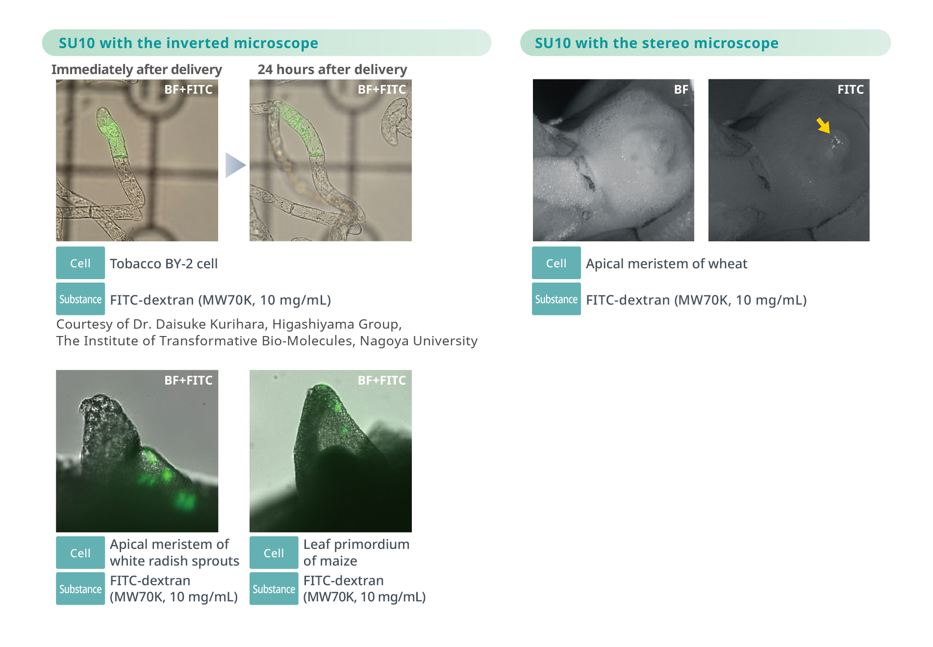
(2) Efficient Delivery into Cultured Plant Cells and Whole Organisms
(3) Efficient Delivery into Challenging Cell Types and Organisms

Publication Information
Featuring a study that utilizes our products.
| Dissertation Title | URL | |
|---|---|---|
| 1 | Electroosmotic Flow-Based Nanoinjection Technique Using a Nanopipette for Green Microalgae. Tanaka T, Akasaka K, Yasui R, Shinohara N, Yoshino T, Nojima D, Mochizuki M, Ohata T, Kamachi F, Sawai T. Mar Biotechnol. 2025 Jul 1;27(4):108. doi: 10.1007/s10126-025-10487-0. |
https://pubmed.ncbi.nlm.nih.gov/40591067/ |
| 2 | Establishment of immortalized murine cardiac fibroblasts for visualizing cytosolic ATP dynamics with a genetically encoded optical indicator Chuluun-Erdene A, Kuchimaru T, Isagawa T, Sato T, Sugimoto H, Ono K, Sawaki D, Sato S, Yamamoto M, Takeda N. Biochem Biophys Res Commun. 2026 Mar 19;805:153377. doi:10.1016/j.bbrc.2026.153377. |
https://www.sciencedirect.com/science/article/abs/pii/S0006291X26001415 |
Specification
| Power voltage | Power voltage | 100 to 240V AC |
|---|---|---|
| Power frequency | 50/60Hz | |
| Power consumption | 70 VA | |
| External dimensions and weight | Main unit | 337 to 456(W)mm×230(H)mm×377(D)mm, 7.4kg |
| Main controller | 133(W)mm×309(H)mm×364(D)mm, 5.9kg | |
| Joystick controller | 140(W)mm×114(H)mm×144(D)mm, 1.2kg | |
| nanopipette | Tip outer diameter : Several tens of nanometers or less (reference value) (in case of SU10ACC-NP02) |
|
| Operation Environment | For use with an inverted optical microscope. * Microscope is not included with the SU10. Please contact Yokogawa to possibly install the SU10 on a different inverted microscope. Installation examples : Nikon Ti2, Evident IX83, Zeiss Axio Observer |
|
Yokogawa Life Science
We post our information on the following social media channels.
Please follow us.
| Yokogawa Life Science | |
| •Ⅹ | @Yokogawa_LS |
| Yokogawa Life Science | |
| •YouTube | Life Science Yokogawa |
Yokogawa's Official Social Media Account List
Ressources

SU10 is a novel technology that enables the delivery of target substances directly into cells (nucleus or cytoplasm) using a "nano" pipette made of a glass capillary with an outer tip diameter of tens of nanometers.
Downloads
Brochures
- Single Cellome Unit SU10 Bulletin (11.2 MB)
Vidéos

YOKOGAWA will contribute to technology evolution particularly in measurement and analytical tools to help build a world where researchers will increasingly focus on insightful interpretation of data, and advancing Life Science to benefit humanity.
Communiqués de presse
-
Communiqué de Presse | Solutions & Products Dec 1, 2021 Yokogawa Develops Single Cellome System SS2000 for Subcellular Sampling
A single-cell analysis solution that revolutionizes efficiency in drug discovery research by automating the collection of specific cells and intracellular components
-
Communiqué de Presse | Solutions & Products Mar 18, 2020 Yokogawa Releases SU10 Single Cellome Unit for Use in Biological Research
- For the creation of a smart cell industry
Rechercher plus d'informations sur nos compétences, technologies et solutions
Contactez-nous