
CV8000 features a cell incubator with an improved airtight design that facilitates the observation of cell behavior over long periods of time. In addition, CV8000 comes with CellPathfinder, a new program that can analyze images of unlabeled cells and 3D images of samples. With these features, CV8000 improves the efficiency of drug discovery research and biomedical research on leading-edge subjects such as iPS and ES cells.
High-speed multi-channel imaging
- Enables high-throughput imaging using up to four cameras and high-power lasers
Live-Cell Assays
- Enables screening using live cells
- Allows observation of dynamic cellular behavior
3D Sample Imaging
- Ideal for observation of organoids and spheroids
- Well suited for MPS (Microphysiological Systems) and related models
Label-free analysis
- Combined with analysis software, enables reliable label-free data acquisition, such as cell and nucleus recognition within brightfield images
High Resolution
- Enables observation of fine intracellular structures such as organelles
Kinetics assays
- Supports high-speed response assays such as Ca2+ flux using drug-dispensing robotic pipetters
Details
State-of-the-art technology that enables you to do what you want
Observe cells as they are -Dual spinning disk confocal system-
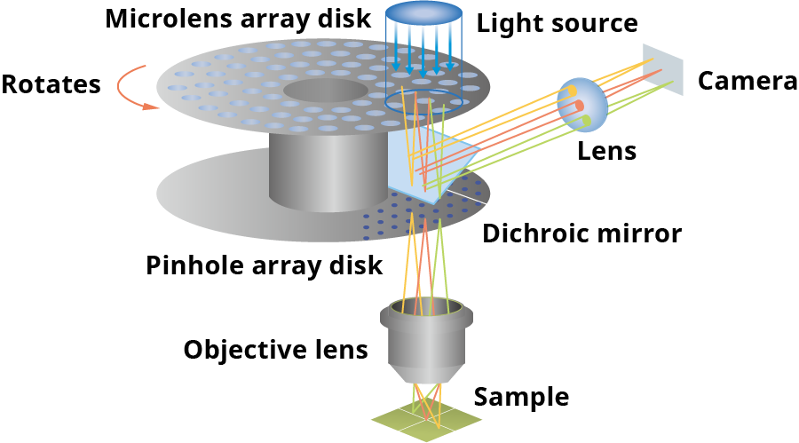
A Yokogawa proprietary multi-scan method utilizing approximately 1,000 laser beams on the observation region and tandem disks rotating at high speed. The disks comprise a pinhole array disk with approximately 20,000 pinholes arranged in an equal pitch spiral pattern, and a microlens array disk that focuses the excitation light laser into individual pinholes. Not only does this allow high speed imaging, but it also largely prevents phototoxicity and fluorescence photobleaching.
Deeper, clearer observation -Pinhole disk exchanger-


Two different types of pinhole disks (25/50μm) can be used, according to the sample. For thick samples, reducing the pinhole diameter allows for higher confocality, shaper images. For dark samples, increasing the pinhole diameter allows for brighter images.
Organoid imaging example Upper:25μm pinhole Lower:50μm pinhole
Higher throughput screening -Optical configuration-

The optical system configuration can be selected according to the purpose. A single 96-well plate can be imaged in four colors in one minute by attaching four high- sensitivity wide -field sCMOS cameras. The system is also compatible with FRET and CellPainting assay.
Capturing finer structures -Original water immersion lens-

Water immersion lenses excel in capturing high-resolution images of cells within a liquid. CV8000 can be equipped with 20x, 40x or 60x water immersion objective lens. Our 40x lens is a particularly unique lens capable of highly advanced spherical aberration correction, allowing for the capture of bright high-resolution images over an entire field of view. The lens water supply is also completely automated. This equipment makes high throughput screening via water submersion lens possible.
Capturing live cell movement -High-precision incubator and robot pipetter-

The stage incubator features an airtight construction, managing humidity, temperature and CO2 levels. The integrated robotic pipetter conducts the following process fully automatically: tip pickup → reagent collection from the reagent plate → reagent addition to the sample plate → tip disposal. Not only can images be rapidly obtained before and after reagent instillation, but it’s also possible to add reagents to single wells multiple times, and adjust the addition speed etc., broadening the range of dynamics observation via reagent instillation.
Application Example
High-resolution images obtained using a water-immersion objective lens equipped with an automatic water supply mechanism provide rich information on drug mechanisms of action and disease pathways. High-speed imaging enabled by Yokogawa's proprietary microlens-equipped Nipkow disk confocal scanner allows the acquisition of high-quality data at high throughput.

Data courtesy of: Charles-Hughes Lardeau & Guy Williams, AstraZeneca, Biopharmaceuticals R&D organization.
A more live cell-friendly total HCA system
Making long-duration live -cell imaging possible -Featuring as table built -in stage incub ator-
HeLa cells were seeded in a 96 well plate at a density of 500 cells per well, and cultured for 24 hours. The well plate was then placed in the internal stage incubator and cell culturing was conducted for 72 hours, and the total area (hereinafter Total Area) occupied by cells was analyzed. As a result, minimal unevenness in cell multiplication was observed across the 96-wells (excluding the four corner wells) when compared to a regular CO2 incubator.

Cell multiplication comparison with regular CO2 incubator after 72hr incubation(n=3)
- 96 well average: 90
- Average of outermost 36 wells: 81
- Average of 60 wells (excl. outermost): 96
The values represent the following: CV8000 Total Area after 72hrs / Total Area at 0hrs (hereinafter Total Area ratio) / CO2 incubator Total Area ratio x 100.
(Numbers near to 100 me an that cell multiplication was approximately equal for CV8000 and CO2 incubator.)
Cell multiplication near to that of the CO2 incubator was verified, excluding the four corner wells.

Cell multiplication curves for each well of a 96-well plate
- Vertical axis: Total Area
- Horizontal axis: Time (0-72 hours)
Cell multiplication was low in the four corner wells; however, it continued in the other wells.

Total Area ratio after cultivation start (24, 48 and 72 hours) (n=3)
Excluding the four corner wells, even after 72 hours, there were no large differences in cell multiplication.
The low variation in cell multiplication speed across the wells can 24 hours 48 hours 72 hours be seen.
More info Evaluation of cell-culture condition in CV8000’s internal stage incubator
High Content Analysis Software CellPathfinder
The software analyzes image data captured with CV8000, creates graphs and exports various data. Beginner and expert users alike can take full advantage of the software, thanks to an abundance of templates and flexible protocol editing capability. CE bright field and machine-learning functionalities make label-free analysis possible. The new Deep Learning option has also been added, largely improving cell recognition accuracy.
More info High Content Analysis Software CellPathfinder
Specifications
High-throughput Cytological Discovery System
| Model | CV8000 |
|---|---|
| Sample format | Multiple well plate (6, 12, 24, 48, 96, 384, 1536 wells), glass slide |
| Image mode | Confocal mode: max. 4 color simultaneous recording Bright field/phase contrast (10x, 20x for 6, 12, 24 well plates), digital phase contrast (10x, 20x) |
| Excitation wavelength | 405/445/488/561/640 nm, all solid laser, max. 5 lasers 【Option】365 nm LED |
| White light illumination | LED |
| Objectives | Max. 6 lenses are available, automatically switchable Dry: 2x, 4x, 10x, 20x, 40x Water immersion: 20x, 40x, 60x Long working distance: 20x Phase contrast: 10x, 20x |
| Confocal unit | Microlens-enhanced wide-view dual Nipkow disk confocal scanner, 50 μm pinhole 【Option】 25 μm pinhole disk and auto pinhole disk exchanger |
| Camera | Max. 4 cameras |
| Stage incubator | Temperature for incubation : 35-40℃ CO2 supply box (CO2: 5%, forced humidification) |
|
Robot pipetter |
【Option】 Disposable tip type (96tip or 384tip type) |
| Bar code reader | 【Option】 1 or 2 dimension |
| Analysis software | High Content Analysis Software CellPathfinder Granularity, Neurite, Nuclear morphology, Nuclear translocation, Plasma membrane translocation, Machine learning, Label-free analysis, 3D analysis, Deep Learning, etc. |
| Power supply | Measurement unit:AC100-240V, 50/60Hz, 2KVA max Workstation for system control:AC100-240V, 50/60Hz, 1.4KVA max Workstation for data analysis:AC100-240V, 50/60Hz, 950VA max |
Yokogawa Life Science
We post our information on the following social media channels.
Please follow us.
| Yokogawa Life Science | |
| •Ⅹ | @Yokogawa_LS |
| Yokogawa Life Science | |
| •YouTube | Life Science Yokogawa |
Yokogawa's Official Social Media Account List
Resources

single-cell analysis specifically transcription by steroid receptors primarily estrogen and androgen receptors
drug discovery and drug development for an interrelated set of disorders that emanate from type II diabetes and obesity

Since 2010, German: Deutsches Zentrum für Neurodegenerative Erkrankungen (DZNE) has been using CellVoyager™ series as the High-content Analysis system of a highly sophisticated screening platform in DZNE Laboratory Automation Technologies (LAT). The team has been collaborating with a lot of scientists and contributing to science advancement via their excellent platform. Dr. Philip Denner is the leader of LAT.

The CV8000 nuclear translocation analysis software enables the analysis of changes in the localization of signal molecules that transfer between cytoplasm and nuclei, such as proteins. The following is an example of the translocation analysis of NFκB, a transcription factor.


Fluorescent ubiquitination-based cell cycle indicator (Fucci) is a set of fluorescent probes which enables the visualization of cell cycle progression in living cells.


Yokogawa collaborates with scientists in the medical and pharmacology fields to identify best practices for cell painting and high-content screening, thereby enhancing image analysis and reproducibility.


List of Selected Publications : CV8000, CV7000, CV6000
Downloads
Brochures
- CV8000 Bulletin (5.5 MB)
Videos

The CV8000 features a cell incubator with an improved airtight design that facilitates the observation of cell behavior over long periods of time. In addition, the CV8000 comes with CellPathfinder, a new program that can analyze images of unlabeled cells and 3D images of samples. With these features, the CV8000 improves the efficiency of drug discovery research and biomedical research on leading-edge subjects such as iPS and ES cells.

YOKOGAWA will contribute to technology evolution particularly in measurement and analytical tools to help build a world where researchers will increasingly focus on insightful interpretation of data, and advancing Life Science to benefit humanity.

In this webinar, Professor Jonny Sexton discusses a pipeline, developed in the Sexton lab, for the quantitative high-throughput image-based screening of SARS-CoV-2 infection to identify potential antiviral mechanisms and allow selection of appropriate drug combinations to treat COVID-19. This webinar presents evidence that morphological profiling can robustly identify new potential therapeutics against SARS-CoV-2 infection as well as drugs that potentially worsen COVID-19 outcomes.


Are you looking to improve laboratory workflows, data management, and imaging analysis?
This webinar covers the integration of a high-content imager into a modern laboratory automation system and workflows built to utilize it. This on-demand webinar describes the integration topology used in the High Throughput Bioscience Center at St. Jude and the technical challenges that emerged pertaining to data handling and analysis. The webinar addresses the variety of ways we have used our high-content imager in the context of a high throughput screening center, using examples of experiment workflows from recent users.
Key Topics:
- Body Copy:
- Key considerations for integrating a high-content imager into a laboratory robot system
- Methods for interfacing between robots and the imager
- Important considerations for imaging data management and analysis
- How the St. Jude High Throughput Bioscience Center supports the diverse imaging needs of its projects


Visualizing the complex spatiotemporal dynamics of human stem cells as they proliferate and make cell fate decisions is key to improving our understanding of how to robustly engineer differentiated tissues for therapeutic applications.
In this webinar, Dr. Rafael Carazo Salas will describe multicolor, multiday high-content microscopy pipelines that his group has recently developed to visualize the dynamical cell fate changes of human Pluripotent Stem Cells (hPSCs).
Key Topics:
- Visualizing how human Pluripotent Stem Cells (hPSCs) proliferate and undergo early differentiation in vitro, by high content microscopy
- Learning about experimental and computational pipelines that enable monitoring single-cell fate dynamics
- Learning about novel “live” reporters of hPSC cell fate
Speaker
Rafael Carazo Salas, PhD
Professor, School of Cellular and Molecular Medicine
University of Bristol

This webinar highlights Yokogawa’s High Content Solutions, the benchtop confocal CellVoyager CQ1, and CellVoyager CV8000. Utilizing Yokogawa’s dual-wide microlens spinning disk confocal technology, these automated HCA systems provide remarkable image quality while increasing your output. This frees up time to complete other research activities. Also, recent additions to the CSU-W1 confocal upgrade is discussed. The SoRa, a super-resolution solution, and the Uniformizer, an image flattening device. Both of which can be added to the lightpath of your CSU-W1-enhanced microscope.
Agenda:
Introduction to Yokogawa
SoRa for CSU-W1 super-resolution with confocal
Two high content instruments from Yokogawa: The CQ1 and the CV8000
Presenter:
Dan J. Collins, Applications Scientist, Yokogawa Life Science

3D imaging experts from Yokogawa and Insphero have come together to provide helpful tips and tricks on acquiring the best 3D spheroid and organoid imaging. This webinar focuses on sample preparation, imaging, and analysis for both fixed and live cells in High Content Screening assays. The experts also discuss automated tools that can help researchers understand the large volume of data in these High Content Imaging Analysis Systems.
News
-
Press Release | Corporate Dec 3, 2020 Yokogawa and InSphero Enter into Partnership Agreement
- Supporting drug development research through the use of HCA and three-dimensional culture models -
-
Press Release | Solutions & Products Sep 5, 2017 Yokogawa Releases CellVoyager CV8000 High-throughput Cytological Discovery System
Looking for more information on our people, technology and solutions?
Contact Us