Upcoming Events
-
Event | Conference May 25 - 28, 2026 Washington D.C., USAMPS World Summit 2026
Join Yokogawa Life Science at the MPS World Summit 2026 in Washington D.C., USA! Experience live demos of the CellVoyager CQ1 for cutting-edge 3D cell imaging and high-content analysis.
What Are Yokogawa's Life Science Solutions for Research and Medicine?
A portfolio of flexible life science products and solutions for the regenerative medicine, pharmaceutical research, and precision medicine industries.
Yokogawa’s high content analysis systems (HCA) and dual spinning disk technologies (CSU), and Single-Cell Analysis are known for higher product quality, more individualized systems tuning, and better process control. From design to implementation and startup to continuous optimization, Yokogawa has the experience and technology to solve your greatest challenges.
Image-Based Solutions
Image-based solutions for science and pharmaceutical drug development.
Yokogawa’s unique imaging designs and analysis tools improve sensitivity and speed for all your scientific exploration. Our instruments provide high-quality data, for maximized research goals.

Automated Instruments
Automated instruments accelerate research by streamlining routine life science tasks.
Yokogawa's automation tackles experimental overload, freeing researchers for more insightful questions and creative exploration. These systems can accelerate drug discovery, time to market, and scientific publication.

Digital Solutions
Digital solutions to monitor and consolidate complex pharmaceutical manufacturing processes.
Yokogawa's robust information sharing tools transcend conventional research organizations. They foster connections that drive problem-solving efficiency and overall experimental effectiveness.

-
Spinning Disk Confocal CSU
CSU confocal unit, featuring a proprietary dual Nipkow disk, excels in live-cell imaging. Its micro lenses boost excitation efficiency for high-speed, low-toxicity imaging.
-
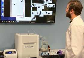
FlowCam: Flow Imaging Microscopy
By analyzing particles accurately, reliably, and quickly via automated imaging, FlowCam® technologies advance research, increase productivity, and ensure quality.
-
Bioprocessing Solutions
Yokogawa’s bioprocessing solutions empower the life sciences industry with integrated automation, real-time analytics, and compliance-ready workflows.
Details
Image-Based Solutions
Image-based solutions for science and pharmaceutical drug development.
Yokogawa’s unique imaging designs and analysis tools improve sensitivity and speed for all your scientific exploration. Our instruments provide high-quality data, for maximized research goals.


Automated Instruments
Automated instruments accelerate research by streamlining routine life science tasks.
Yokogawa's automation tackles experimental overload, freeing researchers for more insightful questions and creative exploration. These systems can accelerate drug discovery, time to market, and scientific publication.


Digital Solutions
Digital solutions to monitor and consolidate complex pharmaceutical manufacturing processes.
Yokogawa's robust information sharing tools transcend conventional research organizations. They foster connections that drive problem-solving efficiency and overall experimental effectiveness.


Yokogawa Life Science
We post our information to the following SNSs.
Please follow us.
| •Ⅹ (former : Twitter) | @Yokogawa_LS |
| Yokogawa Life Science | |
| Yokogawa Life Science | |
| •YouTube | Life Science Yokogawa |
Yokogawa's Official Social Media Account List
Resources

Closing in on Neuronal Circuit Dynamics through High-speed, fMCI.
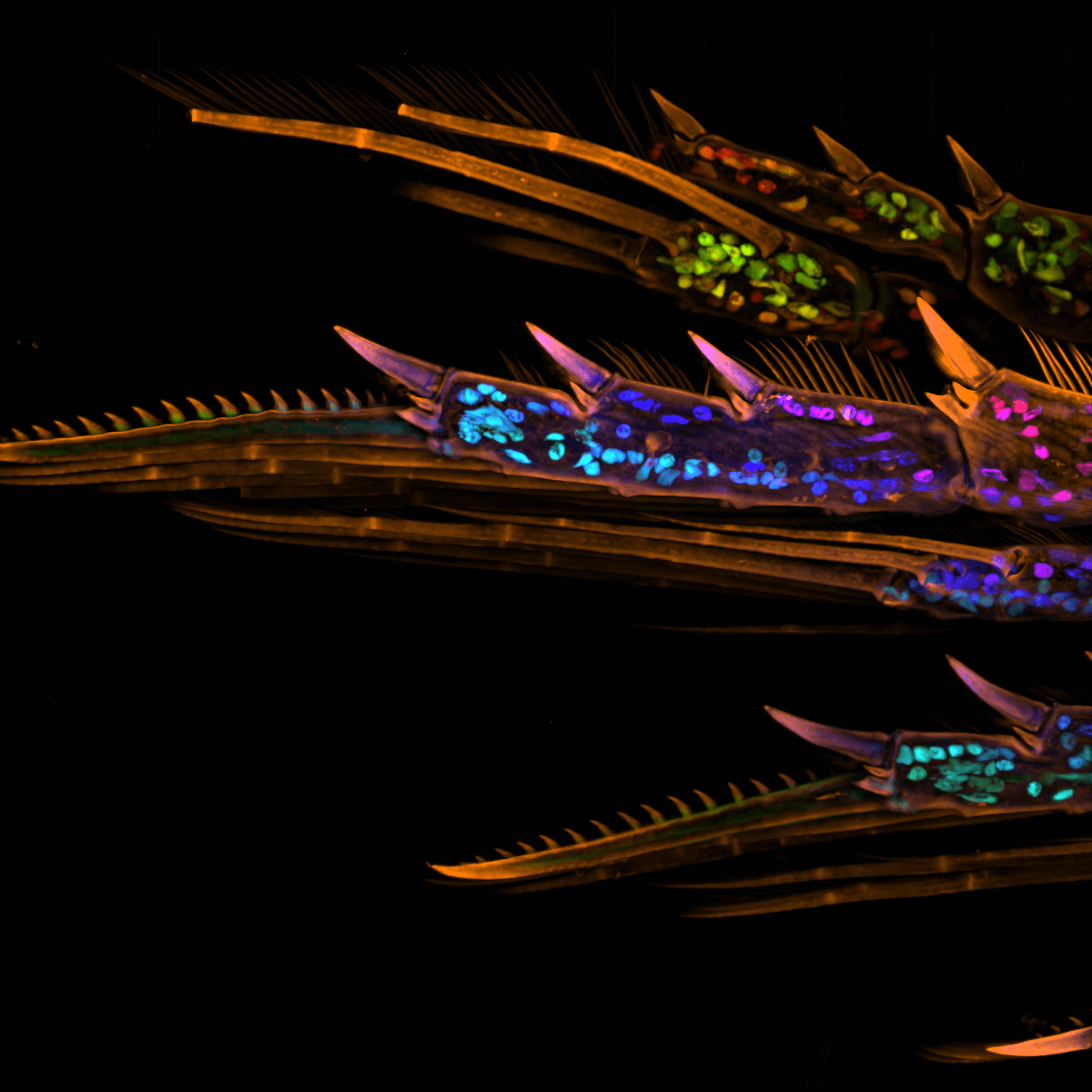
Yokogawa held our annual “Yokogawa CSU Spinning Disk Image Competition.” To highlight the research and advancements of students and scientists who participated in MBL,

First annual Yokogawa CSU Spinning Disk Image Competition at MBL 2023


The SU10 is a “nano” pipette with a tip outer diameter of only a few tens of nanometers, which can automatically deliver target substances into cells (nucleus, cytoplasm) at the single-cell level. The SU10 can directly deliver genome editing tools into the nucleus, so it is expected to enable efficient genome editing. In this application note, we will introduce a case study in which Cas9 RNP and donor DNA were delivered into cells using SU10, resulting in the successful knockout (KO) and knock-in (KI) of target genes.

The CV8000 nuclear translocation analysis software enables the analysis of changes in the localization of signal molecules that transfer between cytoplasm and nuclei, such as proteins. The following is an example of the translocation analysis of NFκB, a transcription factor.


Comparison between CSU and conventional LSM in 4D movies.

To investigate interactive dynamics of the intracellular structures and organelles in the stomatal movement through live imaging technique, a CSU system was used to capture 3-dimensional images (XYZN) and time-laps images (XYT) of guard cells.


Cell stage categorized using FucciTime lapse imaging of Fucci-added Hela cells was conducted over 48 hrs at 1 hr intervals. Gating was performed based on the mean intensities of 488 nm and 561 nm for each cell. They were categorized into four stages, and the cell count for each was calculated.


The CQ1 confocal image acquisition mechanism with the distinctive CSU® unit has a function to sequentially acquire fine cell images along the Z-axis and capture information from the entire thickness of
cells which include heterogenic populations of various cell cycle stages. In addition, saved digital images can be useful for precise observation and analysis of spatial distribution of intracellular molecules.
The CQ1 capability to seamlessly analyze images and obtain data for things such as cell population statistics to individual cell morphology will provide benefits for both basic research and drug discovery
targetingM-cell cycle phase.


Faster, Brighter, and More Versatile Confocal Scanner Unit


High-content screening is a crucial technique in drug development, utilized both in the initial phases for identifying new candidate compounds and in later phases for assessing the pharmacological effects and safety of these candidates. This report highlights three applications using Yokogawa’s CellVoyager CV8000 and CQ1 high-content screening systems, paired with Yokogawa’s CellPathfinder analysis software, creating an integrated and user-friendly platform for drug discovery.
Featured Assays
- Hepatotoxicity Assay: Evaluating the effect of vitamin K on HepG2 cells.
- Cell Migration Assay: Investigating the impact of mitomycin C on cell migration.
- In Vivo Tumor Metastasis: Studying tumor metastasis using zebrafish models.

In this tutorial, a method for analyzing ramified structure, using CellPathfinder, for the analysis of the vascular endothelial cell angiogenesis function will be explained.
In this tutorial, a method for analyzing ramified structure, using CellPathfinder, for the analysis of the vascular endothelial cell angiogenesis function will be explained.

In this tutorial, spheroid diameter and cell (nuclei) count within the spheroid will be analyzed.

In this tutorial, we will learn how to perform time-lapse analysis of objects with little movement using CellPathfinder, through calcium imaging of iPS cell-derived cardiomyocytes.
In this tutorial, we will identify the cell cycles G1-phase, G2/M-phase, etc. using the intranuclear DNA content.

In this tutorial, image analysis of collapsing stress fibers will be performed, and concentration-dependence curves will be drawn for quantitative evaluation.

In this tutorial, we will observe the change in number and length of neurites due to nerve growth factor (NGF) stimulation in PC12 cells.

In this tutorial, intranuclear and intracytoplasmic NFκB will be measured and their ratios calculated, and a dose-response curve will be created.

In this tutorial, we will learn how to perform cell tracking with CellPathfinder through the analysis of test images.

In this tutorial, using images of zebrafish whose blood vessels are labeled with EGFP, tiling of the images and recognition of blood vessels within an arbitrary region will be explained.

Download to learn:
- How spinning-disk confocal innovations improved speed and reduced photodamage
- How the emergence of organoids reshaped experimental design and data requirements
- How advances in 3D imaging, data handling, and analysis enabled large-scale, physiologically relevant drug screening
Downloads
Brochures
- Life Innovation (2.4 MB)
- Yokogawa in the Pharmaceutical Industry (7.7 MB)
- CellVoyager CQ1 Benchtop High-Content Analysis System (2.9 MB)
- FlowCam 8000 Imaging Particle Analysis System (720.4 KB)
Videos

The water and wastewater works around the world have to respond to a wide range of challenges.
Here we introduce Yokogawa‘s three initiatives to address these issues and challenges.


Explore single-cell lipidomics and cell diversity with Yokogawa’s SS2000 in this on-demand webinar. Featuring Kyle Saunders from the University of Surrey’s SEISMIC facility, learn mass spectrometry insights and more—watch now!

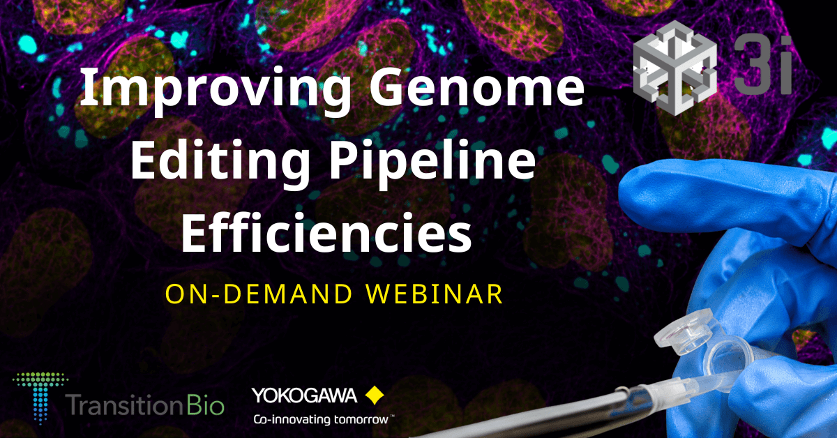
Discover how generative AI and automation are revolutionizing neuroscience drug discovery by enabling scalable, high-throughput human iPSC neuronal models with unprecedented efficiency and translatability, reducing the reliance on less relevant cell lines.
In this webinar, you will learn:
- How to scale and standardize iPSC-derived neuronal models to overcome challenges in complexity, reproducibility, and throughput;
- How integrating iPSC neuronal models early in drug discovery can reduce reliance on less relevant cell lines and improve research translatability;
- How the Neuron Factory platform uses automation, brightfield microscopy, and generative AI to accelerate high-throughput screening and medicinal chemistry workflows.
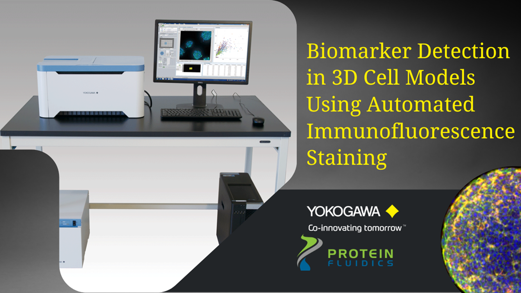
Physiologically relevant 3D cell models are essential for drug discovery and preclinical research due to their functional and architectural similarity to solid tumors. One of the challenges faced by researchers is that many of the assays using these precious samples tend to be manual and tedious.
Using proprietary microfluidics technology, Protein Fluidics has created the Pu·MA System for automated complex 3D cell-based assays. In this webinar, application scientist Dr. Katya Nikolov will present her work on combining this novel automation technology with Yokogawa’s high-content imaging systems for biomarker detection in 3D cell models. Nikolov will demonstrate the utility of an automated immunofluorescence staining workflow followed by confocal imaging within the Pu·MA System flowchips. This automated workflow enables quantitative assessment of biomarkers which provides valuable data for further understanding disease mechanisms, preclinical drug efficacy studies, and in personalized medicine.
This webinar will explore:
- The Pu·MA System and novel technology for automated 3D cell-based assays
- How to perform automated immunofluorescence staining for biomarkers with a “hands-off” assay workflow
- How to visualize biomarkers after the assay with high-content imaging within the flowchip
News
-
News Brief Apr 25, 2024 Yokogawa Helps to Revolutionize the Field of Single-Cell Lipidomics
-
Press Release | Solutions & Products Jan 31, 2024 Yokogawa Introduces CellVoyager High-Content Analysis System CQ3000
- For greater efficiency in drug discovery and regenerative medicine R&D, and the swift commercialization of new drugs -
-
Press Release | Solutions & Products Jun 21, 2023 Yokogawa to Release OpreX Informatics Manager, Enabling Integrated Management of Experimental Data and Research Resources in the Cloud
-
Press Release | Solutions & Products Jun 5, 2020 CSU-W1 Confocal Scanner Unit Arrives at International Space Station
-
Press Release | Corporate Aug 5, 2014 Yokogawa Technology Selected for the Japan Science and Technology Agency's Next-generation Technology Transfer Program
- A major step towards the development of A confocal image single-cell drug discovery support system -
Looking for more information on our people, technology and solutions?
Contact Us