High-Content Analysis Systems
A selection of powerful High-Content Analysis Systems combined with intuitive user software to provide flexible research applications.
Yokogawa’s High Content Analysis Systems (HCA), also known as High Content Screening Systems (HCS), lead the microscopy industry in higher product quality, personalized process control, and streamlined user interfaces. Ideal for advanced analysis tasks, including 3D live cell imaging of spheroids and organoids.
These high-content microscopes, ranging from easy-to-use stand-alone models to fully integrated robotic automation, can be customized to suit diverse needs and workflows, enabling more precise and extensive data collection.
-
CellVoyager High-Content Analysis System CQ3000
CQ3000 can acquire high-resolution 3D images at high speed while culturing cells by combining options according to the application.
-
CV8000 High-Throughput System
Simplify and accelerate workflow processes with the CV8000.
-
Benchtop CQ1 Confocal System
Optimize lab space while driving fast, accurate results.
-
CellPathfinder
Cell imaging software supports label-free samples and provides multiple graphing options with a streamlined, easy-to-use interface.
Details
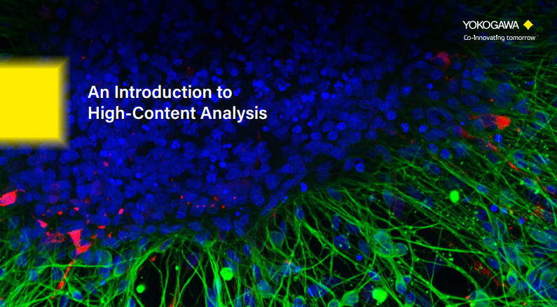
What is High Content Analysis (HCA)?
High-content analysis is a principle methodology for so-called phenotypic screening in drug discovery and development process. Assessing a vast number of compounds is time consuming, therefore, acquiring a large number of high resolution cellular images with fast scanning speed is critically important for drug screening. Yokogawa high-content analysis imagers have balanced these generally conflicting requirements at a high level of performance. The core technology, the microlens-enhanced dual Nipkow disk in Yokogawa Confocal Scanner or Confocal Spinning disc Unit (CSU), makes it possible to capture high quality images with remarkably short acquisition time.
In addition to the image acquisition unit, a precisely controlled fast moving automated stage chamber is also an essential component of our imaging systems. Monolayer fixed cell samples have been commonly used in drug screening. However, cellular environments in such fixed 2D samples are quite different from those of actual live systems, and the divergence often causes a significant discrepancy between experimental results and what is observed in real organisms.
These days, using 3D samples such as spheroids and organoids, and live cell samples to mimic realistic cellular environments are becoming more and more popular due to cell culture technology development. Yokogawa CSU is renowned in the microscopy field not only for the high image quality and fast capturing speed, but also for the live cell friendliness. Our high-content confocal imaging systems based on the CSU are the ideal solution for drug screening using elaborated 3D and live cell assay systems as well as 2D fixed samples. User friendly high content analysis software CellPathfinder, with its versatile functionalities, powerfully supports the analysis of various phenotype changes and target reactions. It also has state-of-the-art machine learning and deep learning function for complex sample analysis. All the technologies are packed into the Yokogawa high-content analysis system.
High Content Analysis vs Traditional Methods
| Traditional Methods | High Content Analysis (HCA) | ||
|---|---|---|---|
| High-Throughput Screening (HTS) | Flow Cytometry (FCM) | Microscopy | |
|
Strength:
Weakness:
|
Strength:
Weakness:
|
Strength:
Weakness:
|
Strengths:
|
Confocal Imagers with Powerful Analysis Software
Our high content analysis (HCA) systems are the best solutions for a range of research applications from basic science to drug discovery screening.
High Content Analysis Publication List
 Powered by Bioz
Resources
PhenoVista Biosciences is the leading provider of custom, imaging-based, phenotypic assay services. With a collaborative and scientifically driven project design and management approach, PhenoVista has a proven track record of delivering high-quality data from robust and scalable assays. PhenoVista’s key advantage lies in the ability of their industry-trained scientists to combine world-class understanding of diverse biological systems with cutting-edge quantitative imaging to deliver clear, actionable output data.

single-cell analysis specifically transcription by steroid receptors primarily estrogen and androgen receptors
drug discovery and drug development for an interrelated set of disorders that emanate from type II diabetes and obesity


The primary goal of high-content imaging is to extract more data from biological samples, including, but not limited to, size, shape, number, and intensity. Here, we describe how the use of Yokogawa’s CQ1 high-content imaging system with microlens-enhanced dual Nipkow disk-based spinning disk confocal, combined with SUN bioscience’s Gri3D® microwell plates, synergize to propel organoid imaging to new heights. Learn more now!

With 462 million affected individuals, diabetes and associated conditions consume about 12% of global health expenditure. Current treatments lack focus on the root cause—the loss of pancreatic insulin-producing beta-cells. This Michigan research team developed a robust, intact ex vivo pancreatic islet bioassay capable of detecting diabetes-relevant endpoints including beta-cell proliferation, chemoprotection, and islet spatial morphometrics.


Cell stage categorized using FucciTime lapse imaging of Fucci-added Hela cells was conducted over 48 hrs at 1 hr intervals. Gating was performed based on the mean intensities of 488 nm and 561 nm for each cell. They were categorized into four stages, and the cell count for each was calculated.


The CV8000 nuclear translocation analysis software enables the analysis of changes in the localization of signal molecules that transfer between cytoplasm and nuclei, such as proteins. The following is an example of the translocation analysis of NFκB, a transcription factor.


Fluorescent ubiquitination-based cell cycle indicator (Fucci) is a set of fluorescent probes which enables the visualization of cell cycle progression in living cells.


Applications: Colony Formation, Scratch Wound, Cytotoxicity, Neurite Outgrowth, Co-culture Analysis, Cell Tracking


The CQ1 confocal image acquisition mechanism with the distinctive CSU® unit has a function to sequentially acquire fine cell images along the Z-axis and capture information from the entire thickness of
cells which include heterogenic populations of various cell cycle stages. In addition, saved digital images can be useful for precise observation and analysis of spatial distribution of intracellular molecules.
The CQ1 capability to seamlessly analyze images and obtain data for things such as cell population statistics to individual cell morphology will provide benefits for both basic research and drug discovery
targetingM-cell cycle phase.

Yokogawa collaborates with scientists in the medical and pharmacology fields to identify best practices for cell painting and high-content screening, thereby enhancing image analysis and reproducibility.


High-content screening is a crucial technique in drug development, utilized both in the initial phases for identifying new candidate compounds and in later phases for assessing the pharmacological effects and safety of these candidates. This report highlights three applications using Yokogawa’s CellVoyager CV8000 and CQ1 high-content screening systems, paired with Yokogawa’s CellPathfinder analysis software, creating an integrated and user-friendly platform for drug discovery.
Featured Assays
- Hepatotoxicity Assay: Evaluating the effect of vitamin K on HepG2 cells.
- Cell Migration Assay: Investigating the impact of mitomycin C on cell migration.
- In Vivo Tumor Metastasis: Studying tumor metastasis using zebrafish models.

This page shows the list of Selected Publications on the Benchtop Confocal System CQ1

List of Selected Publications for High-Content Screening Systems: CV8000, CV7000, CV6000

In this tutorial, we will learn how to perform cell tracking with CellPathfinder through the analysis of test images.

In this tutorial, using images of zebrafish whose blood vessels are labeled with EGFP, tiling of the images and recognition of blood vessels within an arbitrary region will be explained.

In this tutorial, a method for analyzing ramified structure, using CellPathfinder, for the analysis of the vascular endothelial cell angiogenesis function will be explained.

In this tutorial, we will learn how to perform time-lapse analysis of objects with little movement using CellPathfinder, through calcium imaging of iPS cell-derived cardiomyocytes.

In this tutorial, we will observe the change in number and length of neurites due to nerve growth factor (NGF) stimulation in PC12 cells.

In this tutorial, image analysis of collapsing stress fibers will be performed, and concentration-dependence curves will be drawn for quantitative evaluation.
In this tutorial, we will identify the cell cycles G1-phase, G2/M-phase, etc. using the intranuclear DNA content.

In this tutorial, we’ll compare positive and negative condition for the count and total area of lipid droplets by adding the oleic acid or Triacsin C.

In this tutorial, intranuclear and intracytoplasmic NFκB will be measured and their ratios calculated, and a dose-response curve will be created.

In this tutorial, spheroid diameter and cell (nuclei) count within the spheroid will be analyzed.
In this tutorial, a method for analyzing ramified structure, using CellPathfinder, for the analysis of the vascular endothelial cell angiogenesis function will be explained.

Download to learn:
- How spinning-disk confocal innovations improved speed and reduced photodamage
- How the emergence of organoids reshaped experimental design and data requirements
- How advances in 3D imaging, data handling, and analysis enabled large-scale, physiologically relevant drug screening
Downloads
Brochures
Technical Information
Videos

The CV8000 features a cell incubator with an improved airtight design that facilitates the observation of cell behavior over long periods of time. In addition, the CV8000 comes with CellPathfinder, a new program that can analyze images of unlabeled cells and 3D images of samples. With these features, the CV8000 improves the efficiency of drug discovery research and biomedical research on leading-edge subjects such as iPS and ES cells.

Yokogawa's CQ1 open platform integrates seamlessly with Advanced Solutions BioAssemblyBot® 400. With laboratory automation becoming a standard in research, Yokogawa's high content confocal system's ability to work with robots like Advanced Solutions' BioAssemblyBot® 400 is essential to advancing laboratory automation.


Watch the film by KBC who are supporting their clients in their efforts to address climate change and sustainability.
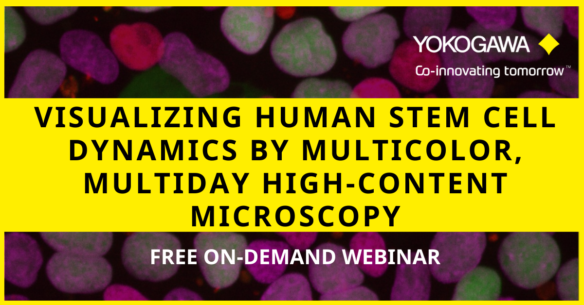
Visualizing the complex spatiotemporal dynamics of human stem cells as they proliferate and make cell fate decisions is key to improving our understanding of how to robustly engineer differentiated tissues for therapeutic applications.
In this webinar, Dr. Rafael Carazo Salas will describe multicolor, multiday high-content microscopy pipelines that his group has recently developed to visualize the dynamical cell fate changes of human Pluripotent Stem Cells (hPSCs).
Key Topics:
- Visualizing how human Pluripotent Stem Cells (hPSCs) proliferate and undergo early differentiation in vitro, by high content microscopy
- Learning about experimental and computational pipelines that enable monitoring single-cell fate dynamics
- Learning about novel “live” reporters of hPSC cell fate
Speaker
Rafael Carazo Salas, PhD
Professor, School of Cellular and Molecular Medicine
University of Bristol
Are you looking to improve laboratory workflows, data management, and imaging analysis?
This webinar covers the integration of a high-content imager into a modern laboratory automation system and workflows built to utilize it. This on-demand webinar describes the integration topology used in the High Throughput Bioscience Center at St. Jude and the technical challenges that emerged pertaining to data handling and analysis. The webinar addresses the variety of ways we have used our high-content imager in the context of a high throughput screening center, using examples of experiment workflows from recent users.
Key Topics:
- Body Copy:
- Key considerations for integrating a high-content imager into a laboratory robot system
- Methods for interfacing between robots and the imager
- Important considerations for imaging data management and analysis
- How the St. Jude High Throughput Bioscience Center supports the diverse imaging needs of its projects
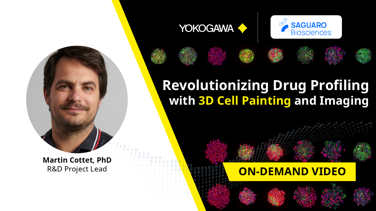
Watch our free on-demand webinar to explore non-toxic 3D cell painting and high-content imaging for enhanced drug profiling and disease phenotyping. Learn how cutting-edge solutions overcome limitations of traditional imaging methods for faster, more accurate research results.


3D imaging experts from Yokogawa and Insphero have come together to provide helpful tips and tricks on acquiring the best 3D spheroid and organoid imaging. This webinar focuses on sample preparation, imaging, and analysis for both fixed and live cells in High Content Screening assays. The experts also discuss automated tools that can help researchers understand the large volume of data in these High Content Imaging Analysis Systems.


Physiologically relevant 3D cell models are being adopted for disease modeling, drug discovery and preclinical research due to their functional and architectural similarity to their tissue/sample of origin, especially for oncology research. Multifunctional profiling and assays using 3D cell models such as tumoroids tend to be manual and tedious. Further, high-content imaging of biomarkers in 3D cell models can be difficult.
In this two-part webinar present to you streamlined technologies which can bring consistent timesaving, ease-of-use, and high-quality data to your 3D cell-based workflows:
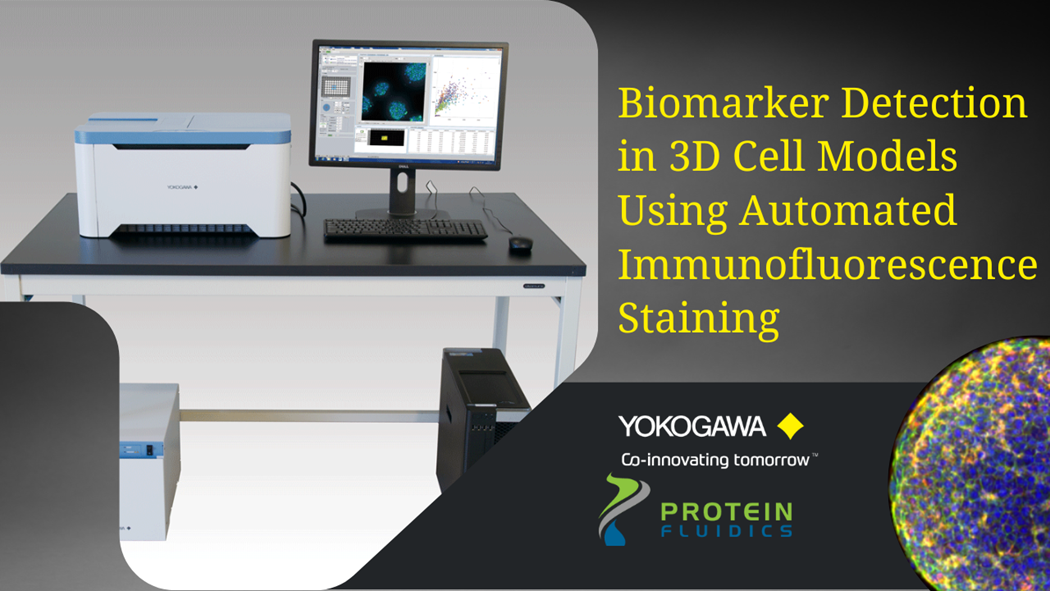
(A) The Pu·MA System is a microfluidics-based benchtop automated device for performing “hands-off” 3D cell-based assays. In this webinar, application scientist Dr. Katya Nikolov will present data from optimized assays using tumoroids followed by Yokogawa’s high-content imaging systems for biomarker detection.
(B) Yokogawa’s high-content imaging systems such as CellVoyager CQ1 provide superior confocal imaging using the Nipkow Spinning Disk Confocal Technology. Here, application scientist, Dan Collins will present details of the high-content imaging capabilities, easy to use and intuitive image acquisition software, especially for increasing productivity and a streamlined workflow.
Learn How:
- The open platform, Pu·MA System can be used to automate your 3D cell-based assays
- To perform automated IF staining for biomarkers using tumoroid models without perturbing your precious samples
- Image acquisition from 3D cell models using Yokogawa’s high-content imaging platforms
- Image analysis from cells, complex spheroids, colonies, or tissues using the CellPathfinder high content analysis software
Physiologically relevant 3D cell models are essential for drug discovery and preclinical research due to their functional and architectural similarity to solid tumors. One of the challenges faced by researchers is that many of the assays using these precious samples tend to be manual and tedious.
Using proprietary microfluidics technology, Protein Fluidics has created the Pu·MA System for automated complex 3D cell-based assays. In this webinar, application scientist Dr. Katya Nikolov will present her work on combining this novel automation technology with Yokogawa’s high-content imaging systems for biomarker detection in 3D cell models. Nikolov will demonstrate the utility of an automated immunofluorescence staining workflow followed by confocal imaging within the Pu·MA System flowchips. This automated workflow enables quantitative assessment of biomarkers which provides valuable data for further understanding disease mechanisms, preclinical drug efficacy studies, and in personalized medicine.
This webinar will explore:
- The Pu·MA System and novel technology for automated 3D cell-based assays
- How to perform automated immunofluorescence staining for biomarkers with a “hands-off” assay workflow
- How to visualize biomarkers after the assay with high-content imaging within the flowchip

This webinar highlights Yokogawa’s High Content Solutions, the benchtop confocal CellVoyager CQ1, and CellVoyager CV8000. Utilizing Yokogawa’s dual-wide microlens spinning disk confocal technology, these automated HCA systems provide remarkable image quality while increasing your output. This frees up time to complete other research activities. Also, recent additions to the CSU-W1 confocal upgrade is discussed. The SoRa, a super-resolution solution, and the Uniformizer, an image flattening device. Both of which can be added to the lightpath of your CSU-W1-enhanced microscope.
Agenda:
Introduction to Yokogawa
SoRa for CSU-W1 super-resolution with confocal
Two high content instruments from Yokogawa: The CQ1 and the CV8000
Presenter:
Dan J. Collins, Applications Scientist, Yokogawa Life Science

In the last few decades, the pharmaceutical industry has transformed people’s lives. However, the development of new drugs is becoming increasingly difficult and a paradigm shift in the drug discovery workflow is required to reduce attrition and transform conventional drug screening assays into translatable analytical techniques for the analysis of drugs in complex environments, both in-vitro and ex-vivo. The ability to visualize unlabelled compounds inside the cell at physiological dosages can offer valuable insight into the compound behavior both on and off-target.
SiLC-MS is a semi-automated methodology that allows the collection of intracellular contents using a modified CQ1 imaging system developed by Yokowaga. The instrument is equipped with a confocal microscope that allows bright field imaging as well as fluorescence imaging with 4 lasers (405, 488, 561, and 640 nm). Sampling is performed using the tips developed by Professor Masujima (1-4).
In this study, we show the applicability of the SiLC-MS technology to drug discovery, as it is crucial to identify compound and its metabolites when incubated in a mammalian cell at a therapeutic dose. We report on the validation studies performed using the SiLC-MS platform, in these validation studies we assess the ability to distinguish different cell types based on their metabolomic fingerprint, furthermore, we have also evaluated if this assay was sensitive enough to detect drugs intracellularly.
Presenter: Carla Newman, Scientific Leader (Celluar Imaging and Dynamics), GSK

Human pluripotent stem cells are proven efficient models for drug screening campaigns. They provide access to unlimited starting material amenable to high throughput small molecule compound screening. Due to their capacity to generate mode complex cellular models, they also offer the potential to perform high content screening in tissue-specific organoids for further human target validation.
Dr. Alejandro Hidalgo-Gonzalez at MCRI(Murdoch Children's Research Institute) in Australia is a user of our HCA system CellVoyager CV8000 and he has established a pipeline for assay development and automated unbiased phenotypic drug screening using human pluripotent stem cell-derived cells and organoids. This is a recording of his presentation at an educational webinar organized by A*STAR in Singapore.
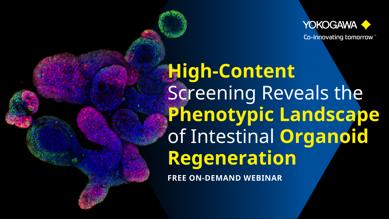
Image-based phenotypic screening relies on the extraction of multivariate information from cells cultured in a large number of screened conditions. In this webinar, we explored the application of complex and biologically relevant model systems for drug screening, such as small intestinal organoids.
Key topics include:
- Learn how to upscale, streamline, and automate intestinal organoid handling
- Learn how to image in complex three-dimensional (3D) model systems and how to approach large imaging datasets
- Understand the basics of multivariate analysis on image-inferred features

Generating translatable high-content imaging data from physiologically-relevant cell models, including 2D and 3D structures, is extremely valuable for drug discovery and pre-clinical research. In this webinar, James Evans, CEO of PhenoVista Biosciences presents case studies on how Yokogawa’s Benchtop CQ1 Confocal System can improve throughput and standardize processes for complex 3D cell-based phenotypic assays.
Key learning objectives:
- Strategies for designing and implementing high-content screening assays
- Approaches for deciding between 2D and 3D model systems
News
-
Press Release | Solutions & Products Jan 31, 2024 Yokogawa Introduces CellVoyager High-Content Analysis System CQ3000
- For greater efficiency in drug discovery and regenerative medicine R&D, and the swift commercialization of new drugs -
-
Press Release | Solutions & Products Nov 30, 2021 Yokogawa Develops Single Cellome System SS2000 for Subcellular Sampling
Looking for more information on our people, technology and solutions?
Contact Us