What is Dual Spinning Disk Confocal Technology?
As the pioneer in dual-spinning disk confocal technology, we have revolutionized live-cell imaging in optical microscopy. The multi-beam scanning method offers not only high-speed imaging but also significantly reduced phototoxicity and photo bleaching, making our confocal scanner units the de facto standard tool for live-cell imaging.
-
Super Resolution
Optimize results with the CSU-W1 SoRa Confocal Scanner.
-
Wide Field of View
Improve efficiency with the CSU-W1 Confocal Scanner.
-
High Speed
Get next level turnarounds with the CSU-X1 Confocal Scanner.
-
Uniformizer
A flat-top beam shaper option for CSU-W1.
Details
High-Speed, High Resolution Imaging
Yokogawa’s confocal scanners utilize advanced imaging technologies to enable researchers to achieve high-speed and high-resolution live cell imaging.
- Fast time-lapse confocal images of living cells
- Minimal phototoxicity and less photobleaching
- Live-cell confocal fluorescence imaging capabilities
- Stability during long-term and high-speed imaging
- Facilitates quantitative analysis of huge amounts of data
Comparison of CSU Series
| Model | CSU-W1 | CSU-X1 | ||||
|---|---|---|---|---|---|---|
| High-end | Basic | |||||
| Imaging Speed (Max. fps) |
200 | 2,000 | 360 | |||
| Scanner Motor Rotation Speed (rpm) |
1,500-4,000 | 1,500-10,000 (Variable) |
1,800 (Fixed)*2 |
|||
| Recommended camera exposure time |
5msec | 0.5msec | 33msec | |||
| Effective FOV | 17x16mm | 10x7mm | ||||
| Disk unit | Selectable up to 2 disks Pinhole size : 50µm, 25µm |
1 disk Pinhole size : 50µm |
||||
| Rotation position trigger signal |
External signal output possible | None*2 | ||||
| Filters | EX | Option | ||||
| DM | Option (up to 3 filters) | Option (1 filter) |
||||
| EM | Option (up to 10 filters with filter wheel) |
Option (up to 12 filters with filter wheel) |
Option (1 filter) |
|||
| Addition or exchange of filters |
At user site : DM block and filters (EX, EM.) At Yokogawa factory : DM |
|||||
| *1 option | ||||||
Comparison between CSU and other confocal systems
| Model | CSU | Conventional Point-scan Confocal |
Conventional Slit-scan Confocal |
Other spinning disk Confocal |
Epi-fluorescence (Wide field) |
|---|---|---|---|---|---|
| Scan Type | Microlens-enhanced multi beam scan |
Single beam scan | Line Scan | Disk scan (Multi beam or slit) | None |
| Light Source | Lasers | Hg or Xenon arc lamp | |||
| Detector | CCD, EMCCD | PMT | Line CCD | CCD, EMCCD | |
| Microscope | Flexible | Specific | Flexible / Specific | Flexible | |
| Scan speed of full-size image |
2000fps | ~1fps | ~120fps (512X512) | <200fps | Any |
| Photo bleaching/ photo toxicity |
Low | Severe | Low | ||
| Confocality | High (X-Y-Z confocal) | Modest (Compromise y-resolution) |
Modest (X-Y-Z confocal with pinhole, Compromise y-resolution with slit-scan) |
None | |
| Image Quality (Background) |
High Good S/N (Low background) |
High (multiple averaging necessary) |
Modest | Modest (High background with dim samples) |
Low (High background) |
User labs
- Ted Salmon Lab., Dept. of Biology, University of North Carolina, Chapel Hill
- Waterman-Storer Lab., Laboratory of Cell and Tissue Morphodynamics (LCTM),NHLBI (Bethesda Campus)
- Tim Mitchison Lab., Dept. of Systems Biology, Harvard Medical School
- Scholey Lab., Dept. of Cell and Computational Biology, University of California, Davis
- The Vale Lab., Dept. of Cellular and Molecular Pharmacology, University of California, San Francisco
- The Wadsworth Lab., Biology Dept., University of Massachusetts, Amherst
- The Kiehart Lab., Dept. of Biology, Duke University
- HSC Core Facilities School of Medicine, University of Utah
- Indiana Center for Biological Microscopy, Indiana University Medical Center
- Ehlers Laboratory - Department of Neurobiology, Duke University
- Bob Goldstein Lab., University of North Carolina Chapel Hill
- Andrew Matus Lab., at Friedrich Miescher Institute for Biomedical Research
- Laboratory of Developmental Dynamics,Graduate School of Life Sciences, Tohoku University
- Zena Werb Lab., Anatomy, University of California San Francisco
- Satoshi Nishimura Lab., Dept. of Cardiovascular Medicine, the University of Tokyo
- Oshima Lab., Graduate School of Interdisciplinary Information Studies,The Univ. of Tokyo
- The Huser research group at UC Davis
- Nakano Lab., Graduate School of Science, University of Tokyo
Sites and Textbooks
1) Microscopy & Imaging Resources on the WWW
Complete list of all aspects of microscopy and imaging, by Douglas W. Cromey From Cellular Imaging Core of Southwest Environmental Health Sciences Center, University of Arizona College of Pharmacy, University of Arizona
2) The Centro de Biologia Molecular “Severo Ochoa” (CBMSO)
Contains links to general information, microscopy laboratories, publications, courses and meetings, societies, images. Especially useful for finding microscopy workshops.
3) The Cell Imaging Facility, a part of the University of Utah Health Science Center's Core Research Facilities Department
Good explanation of CSU-Disk scanning confocal system by Chris Rodesch
4) Molecular Expressions Website
Run by National High Magnetic Field Laboratory, Florida State University.
One of Web's largest collections of excellent optical microscopy images, and quite thorough information on all types of microscopy.
Interactive Java Tutorials sponsored by Nikon (Nikon MicroscopyU) and Olympus(Olympus Microscopy Resource Center) are extremely useful to learn not only confocal but all kinds of microscopies, and related technologies.
Life Science Textbooks
|
Microscopy Techniques, Advances in Biochemical Engineering / Biotechnology Vol.95 |
|
|
Confocal Microscopy for Biologists |
|
|
Live Cell Imaging, A Laboratory Manual |
|
|
Handbook of Biological Confocal Microscopy, 3rd Edition |
|
|
VideoMicroscopy, The Fundamentals ISBN: 0-306-45531-5
|
|
|
Direct-View High-Speed Confocal Scanner: The CSU-10, Chapter 2: Cell Biological Applications of Confocal Microscopy (Methods in Cell Biology) |
Spinning Disk Confocal Publication List
 Powered by Bioz
Resources

Discovering the Basic Principles of Life through the Live Imaging of C. elegans

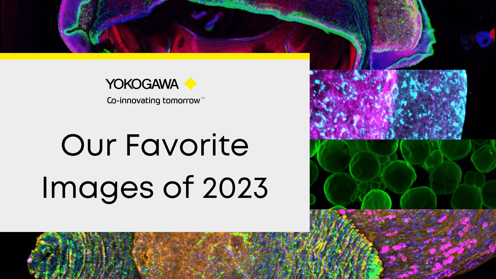
First annual Yokogawa CSU Spinning Disk Image Competition at MBL 2023
Visualizing the cell behavioral basis of epithelial morphogenesis and epithelial cancer progression

Spinning Disk Confocal Microscopy for Quantitative Imaging and Multi-Point Fluorescence Fluctuation Spectroscopy.


Comparison between CSU and conventional LSM in 4D movies.


To investigate interactive dynamics of the intracellular structures and organelles in the stomatal movement through live imaging technique, a CSU system was used to capture 3-dimensional images (XYZN) and time-laps images (XYT) of guard cells.


Faster, Brighter, and More Versatile Confocal Scanner Unit


List of Selected Publications for the High-Speed Confocal Scanner (CSU-X1)

List of Selected Publications for the Wide Field of View Confocal Scanner (CSU-W1)
Downloads
Brochures
- CSU-W1 Confocal Scanner Unit (2.9 MB)
- CSU-X1 Confocal Scanner Unit (2.4 MB)
- Yokogawa CSU-W1 SoRa Confocal Scanner Unit (525.0 KB)
Videos

YOKOGAWA proprietary Spinning Disk technology enables fast real-time confocal imaging for applications such as high-speed 3D and long-term live cell imaging. These quantifiable imaging analysis are essential tools for modern precision drug discovery.

Fast, gentle, and clear - live-cell imaging. Yokogawa's unique scanning method minimizes damage to living cells and organisms and even can capture faint/fast life phenomena.
More than 2,500+ units scanning units sold worldwide. This fast, reliable, and accurate technology has been leading cutting-edge research and supporting researchers around the world for more than two decades.
More information: https://www.yokogawa.com/us/solutions/products-platforms/life-science/spinning-disk-confocal/
#confocal #microlens #microscope #CSU #Yokogawa #livecell

This webinar highlights Yokogawa’s High Content Solutions, the benchtop confocal CellVoyager CQ1, and CellVoyager CV8000. Utilizing Yokogawa’s dual-wide microlens spinning disk confocal technology, these automated HCA systems provide remarkable image quality while increasing your output. This frees up time to complete other research activities. Also, recent additions to the CSU-W1 confocal upgrade is discussed. The SoRa, a super-resolution solution, and the Uniformizer, an image flattening device. Both of which can be added to the lightpath of your CSU-W1-enhanced microscope.
Agenda:
Introduction to Yokogawa
SoRa for CSU-W1 super-resolution with confocal
Two high content instruments from Yokogawa: The CQ1 and the CV8000
Presenter:
Dan J. Collins, Applications Scientist, Yokogawa Life Science

In the last few decades, the pharmaceutical industry has transformed people’s lives. However, the development of new drugs is becoming increasingly difficult and a paradigm shift in the drug discovery workflow is required to reduce attrition and transform conventional drug screening assays into translatable analytical techniques for the analysis of drugs in complex environments, both in-vitro and ex-vivo. The ability to visualize unlabelled compounds inside the cell at physiological dosages can offer valuable insight into the compound behavior both on and off-target.
SiLC-MS is a semi-automated methodology that allows the collection of intracellular contents using a modified CQ1 imaging system developed by Yokowaga. The instrument is equipped with a confocal microscope that allows bright field imaging as well as fluorescence imaging with 4 lasers (405, 488, 561, and 640 nm). Sampling is performed using the tips developed by Professor Masujima (1-4).
In this study, we show the applicability of the SiLC-MS technology to drug discovery, as it is crucial to identify compound and its metabolites when incubated in a mammalian cell at a therapeutic dose. We report on the validation studies performed using the SiLC-MS platform, in these validation studies we assess the ability to distinguish different cell types based on their metabolomic fingerprint, furthermore, we have also evaluated if this assay was sensitive enough to detect drugs intracellularly.
Presenter: Carla Newman, Scientific Leader (Celluar Imaging and Dynamics), GSK
News
-
Press Release | Solutions & Products Nov 30, 2021 Yokogawa Develops Single Cellome System SS2000 for Subcellular Sampling
Looking for more information on our people, technology and solutions?
Contact Us